. 2013;8(2):e56305.
doi: 10.1371/journal.pone.0056305. Epub 2013 Feb 14.

Phylogenetic analysis of glucosyltransferases and implications for the coevolution of mutans streptococci with their mammalian hosts

Affiliations

Phylogenetic analysis of glucosyltransferases and implications for the coevolution of mutans streptococci with their mammalian hosts

Silvia Argimón et al. PLoS One. 2013.

Abstract

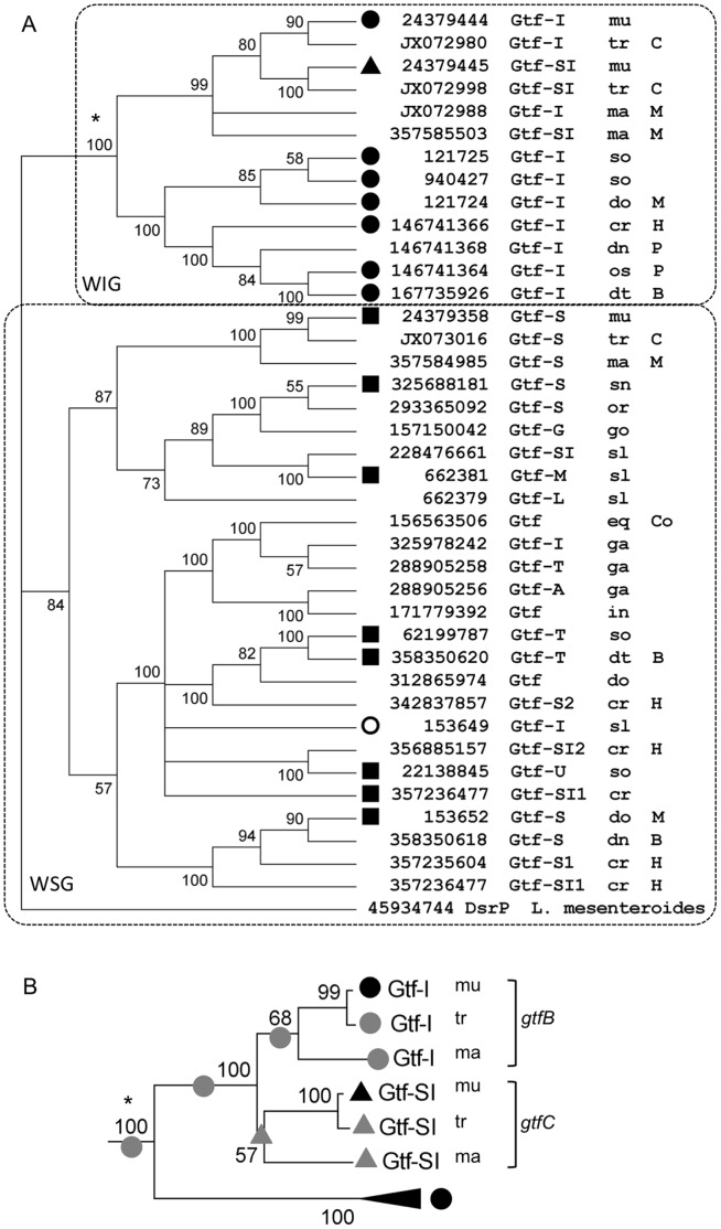
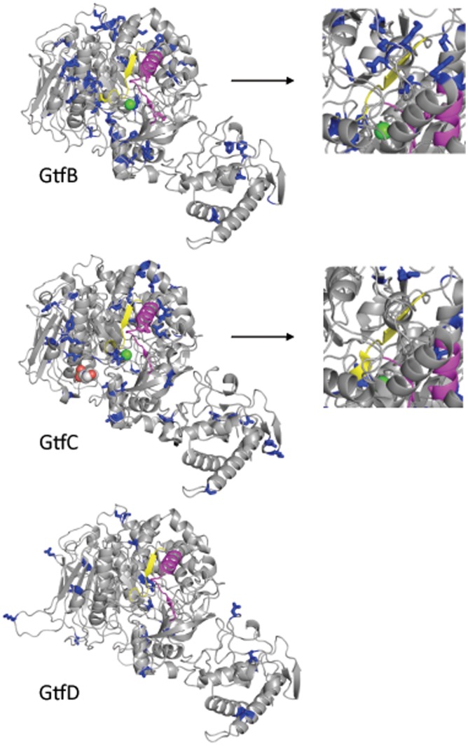
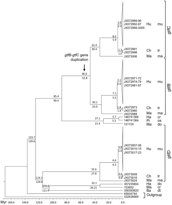
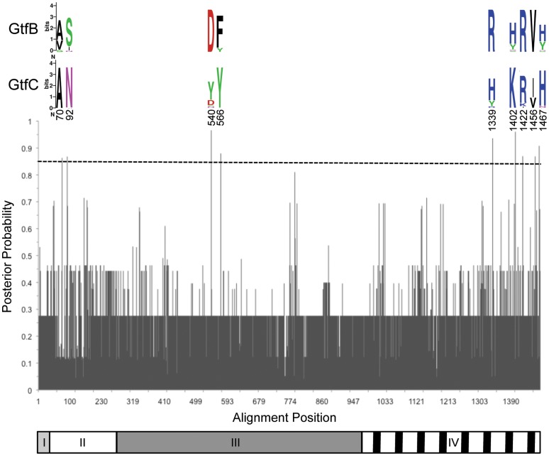
Glucosyltransferases (Gtfs) catalyze the synthesis of glucans from sucrose and are produced by several species of lactic-acid bacteria. The oral bacterium Streptococcus mutans produces large amounts of glucans through the action of three Gtfs. GtfD produces water-soluble glucan (WSG), GtfB synthesizes water-insoluble glucans (WIG) and GtfC produces mainly WIG but also WSG. These enzymes, especially those synthesizing WIG, are of particular interest because of their role in the formation of dental plaque, an environment where S. mutans can thrive and produce lactic acid, promoting the formation of dental caries. We sequenced the gtfB, gtfC and gtfD genes from several mutans streptococcal strains isolated from the oral cavity of humans and searched for their homologues in strains isolated from chimpanzees and macaque monkeys. The sequence data were analyzed in conjunction with the available Gtf sequences from other bacteria in the genera Streptococcus, Lactobacillus and Leuconostoc to gain insights into the evolutionary history of this family of enzymes, with a particular emphasis on S. mutans Gtfs. Our analyses indicate that streptococcal Gtfs arose from a common ancestral progenitor gene, and that they expanded to form two clades according to the type of glucan they synthesize. We also show that the clade of streptococcal Gtfs synthesizing WIG appeared shortly after the divergence of viviparous, dentate mammals, which potentially contributed to the formation of dental plaque and the establishment of several streptococci in the oral cavity. The two S. mutans Gtfs capable of WIG synthesis, GtfB and GtfC, are likely the product of a gene duplication event. We dated this event to coincide with the divergence of the genomes of ancestral early primates. Thus, the acquisition and diversification of S. mutans Gtfs predates modern humans and is unrelated to the increase in dietary sucrose consumption.


Conflict of interest statement

Competing Interests: The authors have declared that no competing interests exist.

Figures

Figure 1
Figure 1. Phylogenetic analysis of streptococcal glucosyltransferases.
A) Maximum likelihood consensus tree of 39 streptococcal glucosyltransferases based on the amino acid sequence of the catalytic domain. Node values indicate bootstrap support from 500 replicates. Branches with less than 50% support were collapsed. Inset: tree topology for the S. mutans water-insoluble glucan cluster based on the full length amino acid sequence. Biochemical data on the type of glucan is indicated as circles for WIG, squares for WSG, and triangles for gtfs that synthesize both WIG and WGS. Bacterial species are indicated as follows: cr = S. criceti; dt = S. dentirousetti; dn = S. dentisuis; do = S. downei; eq = S. equinus; ga = S. gallolyticus; go = S. gordonii; in = S. infantarius; ma = S. macacae; mu = S. mutans; or = S. oralis; os = S. orisuis; sl = S. salivarius; sn = S. sanguinis; so = S. sobrinus; tr = S. troglodytae. The strains were isolated from human subjects unless indicated as follows: B = bat; C = chimpanzee; Co = cow; H = hamster; M = macaque; P = pig. B) Detail of the tree topology for the WIG cluster of S. mutans Gtfs based on the full length amino acid sequence. Reconstruction of character states is indicated by gray circles and triangles.
Figure 2
Figure 2. Models of the 3D structure of the catalytic domain of S. mutans Gtfs.
Blue: amino acids under negative selection (side chains shown). Yellow: active site. Magenta: Gtf-P1 region . Green sphere: Ca2+ ion. Right panels: Detail of a different view of the region surrounding the active site of GtfB and GtfC.
Figure 3
Figure 3. Bayesian phylogeny of streptococcal Gtfs.
Values represent mean node ages obtained from either the nucleotide (top) or the amino acid (bottom) sequences, with the node calibration from . The substitution models were TN93+Γ+I and WAG+Γ+I for nucleotide and amino acid data, respectively. The scale at the bottom represents time before present in millions of years. Bacterial species are indicated as follows: cr = S. criceti; dt = S. dentirousetti; do = S. downei; ma = S. macacae; mu = S. mutans; os = S. orisuis; tr = S. troglodytae. Strains isolated from Hu = human; Ch = chimpanzee; Ma = macaque; Ha = hamster; Pi = pig; Ba = bat.
Figure 4
Figure 4. Site specific profile of type I functional divergence posterior probability.
Logos are shown for positions predicted to be critical for type I functional divergence between GtfB and GtfC (cutoff P>0.85). Residues are color coded by biochemical property and heights represent their relative frequency at each site. The Gtf protein domains are represented below the graph. I) signal peptide, II) N-terminal variable region, III) catalytic domain, IV) glucan binding domain.

References

    1. Loesche WJ (1993) Dental Caries: A Treatable Infection. Grand Haven, Michigan USA: ADD Publications.
    1. Schilling KM, Bowen WH (1992) Glucans synthesized in situ in experimental salivary pellicle function as specific binding sites for Streptococcus mutans . Infect Immun 60: 284–295. - PMC - PubMed
    1. Hannig C, Ruggeri A, Al-Khayer B, Schmitz P, Spitzmuller B, et al. (2008) Electron microscopic detection and activity of glucosyltransferase B, C, and D in the in situ formed pellicle. Arch Oral Biol 53: 1003–1010. - PubMed
    1. Vacca-Smith AM, Bowen WH (1998) Binding properties of streptococcal glucosyltransferases for hydroxyapatite, saliva-coated hydroxyapatite, and bacterial surfaces. Arch Oral Biol 43: 103–110. - PubMed
    1. Gregoire S, Xiao J, Silva BB, Gonzalez I, Agidi PS, et al. (2011) Role of glucosyltransferase B in interactions of Candida albicans with Streptococcus mutans and with an experimental pellicle on hydroxyapatite surfaces. Appl Environ Microbiol 77: 6357–6367. - PMC - PubMed

Publication types

Substances

LinkOut - more resources