Comparative Study
. 2013;8(2):e56315.
doi: 10.1371/journal.pone.0056315. Epub 2013 Feb 14.

Cotton fiber cell walls of Gossypium hirsutum and Gossypium barbadense have differences related to loosely-bound xyloglucan

Affiliations
Comparative Study

Cotton fiber cell walls of Gossypium hirsutum and Gossypium barbadense have differences related to loosely-bound xyloglucan

Utku Avci et al. PLoS One. 2013.

Abstract

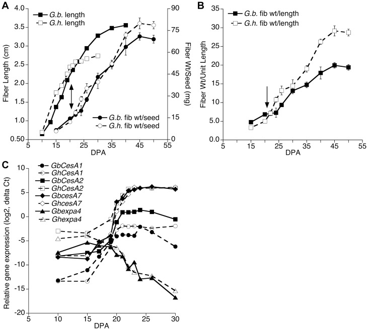
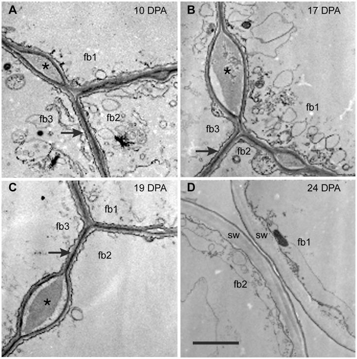
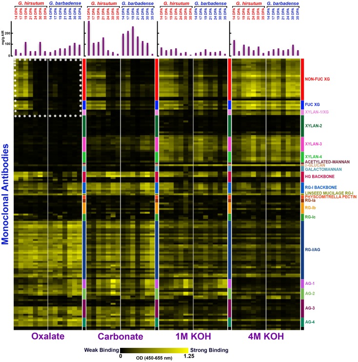
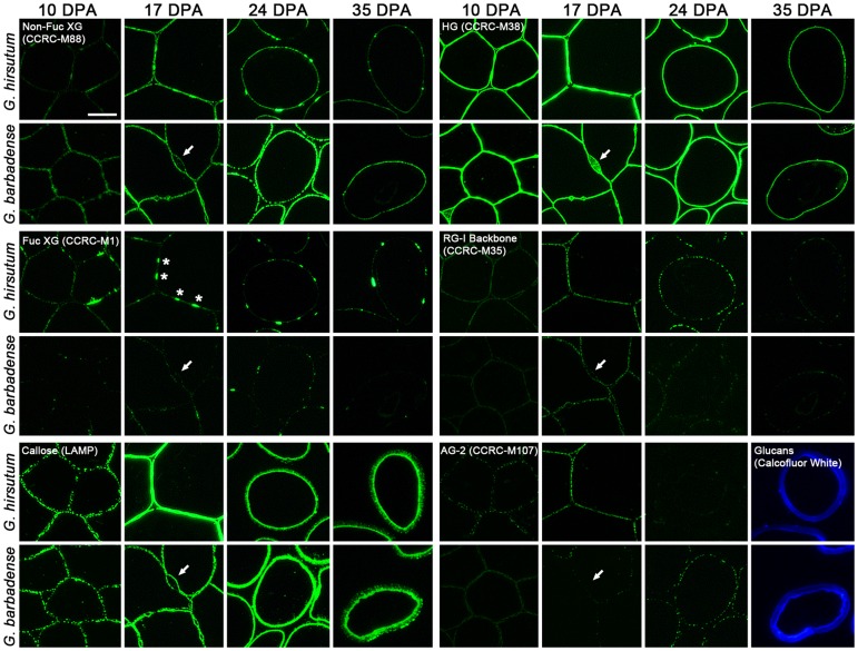
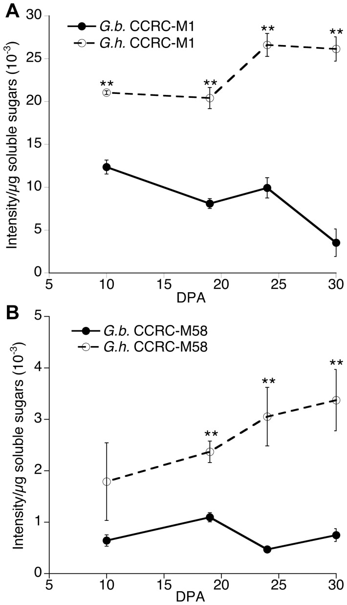
Cotton fiber is an important natural textile fiber due to its exceptional length and thickness. These properties arise largely through primary and secondary cell wall synthesis. The cotton fiber of commerce is a cellulosic secondary wall surrounded by a thin cuticulated primary wall, but there were only sparse details available about the polysaccharides in the fiber cell wall of any cotton species. In addition, Gossypium hirsutum (Gh) fiber was known to have an adhesive cotton fiber middle lamella (CFML) that joins adjacent fibers into tissue-like bundles, but it was unknown whether a CFML existed in other commercially important cotton fibers. We compared the cell wall chemistry over the time course of fiber development in Gh and Gossypium barbadense (Gb), the two most important commercial cotton species, when plants were grown in parallel in a highly controlled greenhouse. Under these growing conditions, the rate of early fiber elongation and the time of onset of secondary wall deposition were similar in fibers of the two species, but as expected the Gb fiber had a prolonged elongation period and developed higher quality compared to Gh fiber. The Gb fibers had a CFML, but it was not directly required for fiber elongation because Gb fiber continued to elongate rapidly after CFML hydrolysis. For both species, fiber at seven ages was extracted with four increasingly strong solvents, followed by analysis of cell wall matrix polysaccharide epitopes using antibody-based Glycome Profiling. Together with immunohistochemistry of fiber cross-sections, the data show that the CFML of Gb fiber contained lower levels of xyloglucan compared to Gh fiber. Xyloglucan endo-hydrolase activity was also higher in Gb fiber. In general, the data provide a rich picture of the similarities and differences in the cell wall structure of the two most important commercial cotton species.


Conflict of interest statement

Competing Interests: The authors have declared that no competing interests exist.

Figures

Figure 1
Figure 1. Fiber developmental progression between 10 to 50 DPA for G. hirsutum (Gh) and G. barbadense (Gb) documented by fiber measurements and gene expression.
(A, B) Fiber length (hand measured) and fiber weight/seed (A) and the ratio of fiber weight to length (B). The timing of visible CFML degradation, which began ∼21 DPA near the onset of secondary wall deposition, is marked by arrows; see the data for Gb in Figure 2 and for Gh published previously (Singh et al., 2009). The data points (± SE) are the means of fiber measurements from 3 to 5 bolls of different plants. (C) Quantitative reverse transcription PCR data showing the developmental shift in gene expression prior to the onset of secondary wall cellulose synthesis at ∼22 DPA. For both G. hirsutum and G. barbadense fiber, the expression of three secondary wall-associated CESA genes began a sharp increase and the expression of an expansin isoforms began a sharp decline at ∼17 DPA. Each data point is the mean from 3 biological replicates. The fiber growth data for Gh up to 32 DPA (A) are republished from Figure S1 of (http://www.plantphysiol.org, Copyright American Society of Plant Biologists).
Figure 2
Figure 2. Transmission electron micrographs of cross-sectioned G. barbadense fiber at 10 to 24 DPA.
The ‘fb#’ labels indicate 2 or 3 individual fibers in each view. (A, B, C) At 10 DPA, 17 DPA, and 19 DPA, adjacent fibers are joined together by the CFML, the outermost layer of the primary wall. In many regions, a thin continuous wall exists between adjacent fibers (arrows in A, B, C). However, there are also periodic bulges between fibers that are filled with CFML material (asterisks in A, B, C). (D) At 24 DPA during secondary wall (sw) deposition, the fibers have separated due to CFML degradation, leaving empty space between them. The 2 µm scale bar in D applies to all micrographs.
Figure 3
Figure 3. Glycome Profiling of sequential extracts prepared from multiple fiber samples representing major stages of cotton fiber development in G. hirsutum and G. barbadense.
A vertical color-coded strip shows the clades of cell wall glycan-binding antibodies (as defined in [21]). Each successive cell wall solvent is shown on the bottom, with a split panel above each label showing the results for Gh and Gb fiber at 14 to 35 DPA. Absorbance values >0.10 typically are perceived as non-black. The bar graphs show the amount of cell wall material removed by each solvent from each sample (mg extracted/g AIR). The dotted white box outlines binding of XG-directed antibodies to the oxalate extracts, where the most striking differences between the two species were observed.
Figure 4
Figure 4. Fluorescence immunohistochemistry of G. hirsutum and G. barbadense fiber cross-sections at 10 to 35 DPA.
The antibodies recognized epitopes found in XG (as shown by CCRC-M88 and CCRC-M1 labeling), ß-1,3-glucan or callose (LAMP), de-esterified HG (CCRC-M38), the RG-I backbone (CCRC-M35), and AG-2 (CCRC-M107). Calcofluor White, which recognizes cellulose and callose, stained the thickened fiber secondary wall at 35 DPA (images substituted for black panels in the lower right). The arrows indicate the location of a bulged region of the CFML in Gb fiber, which showed only HG in its interior. In contrast, HG and XG were in bulged regions of the CFML in Gh fiber (see the asterisks in the CCRC-M1 panel and [3]). Callose was detected within the thickening secondary wall at 35 DPA, whereas the XG and HG epitopes were only in the persistent primary wall on the fiber perimeter. The micrographs for each antibody were taken at the same exposure time. The 10 µm bar in the upper left panel applies to all micrographs.
Figure 5
Figure 5. Colorimetric intensity values from immuno-dot-assays of epitopes within cell wall polymers extracted from G. hirsutum and G. barbadense fiber at 10, 19, 24, and 30 DPA by hot acidic water.
Antibody probes used were: (A) CCRC-M1 recognizing fucosylated XG epitopes and (B) CCRC-M58 recognizing non-fucosylated XG epitopes. Data points are the means (± SE) of 3 biological replications, and asterisks indicate that means from the two species at that DPA are significantly different as determined by T test (**p≤0.01).
Figure 6
Figure 6. Xyloglucan endo-hydrolase (XEH) activity in developing 10 to 30 DPA fibers of G. hirsutum and G. barbadense.
Data points are the means (± SE) of 4 biological replications. The data for Gh are republished from Table SVI of (http://www.plantphysiol.org, Copyright American Society of Plant Biologists).

References

    1. Meinert MC, Delmer DP (1977) Changes in biochemical composition of the cell wall of the cotton fiber during development. Plant Physiol 59: 1088–1097. - PMC - PubMed
    1. Kim HJ, Triplett BA (2001) Cotton fiber growth in planta and in vitro. Models for plant cell elongation and cell wall biogenesis. Plant Physiol 127: 1361–1366. - PMC - PubMed
    1. Singh B, Avci U, Eichler Inwood SE, Grimson MJ, Landgraf J, et al. (2009) A specialized outer layer of the primary cell wall joins elongating cotton fibers into tissue-like bundles. Plant Physiol 150: 684–699. - PMC - PubMed
    1. Haigler CH, Betancur L, Stiff MR, Tuttle JR (2012) Cotton fiber: a powerful single-cell model for cell wall and cellulose research. Front Plant Sci 3: Article 104. Available: http://www.frontiersin.org/Plant_Physiology/10.3389/fpls.2012.00104/full. Accessed 2012 Aug 3. - DOI - PMC - PubMed
    1. Hovav R, Chaudhary B, Udall JA, Flagel L, Wendel JF (2008) Parallel domestication, convergent evolution and duplicated gene recruitment in allopolyploid cotton. Genetics 179: 1725–1733. - PMC - PubMed

Publication types

LinkOut - more resources