. 2013;8(2):e56631.
doi: 10.1371/journal.pone.0056631. Epub 2013 Feb 14.

Bioactivity-guided identification and cell signaling technology to delineate the lactate dehydrogenase A inhibition effects of Spatholobus suberectus on breast cancer

Affiliations

Bioactivity-guided identification and cell signaling technology to delineate the lactate dehydrogenase A inhibition effects of Spatholobus suberectus on breast cancer

Zhiyu Wang et al. PLoS One. 2013.

Abstract

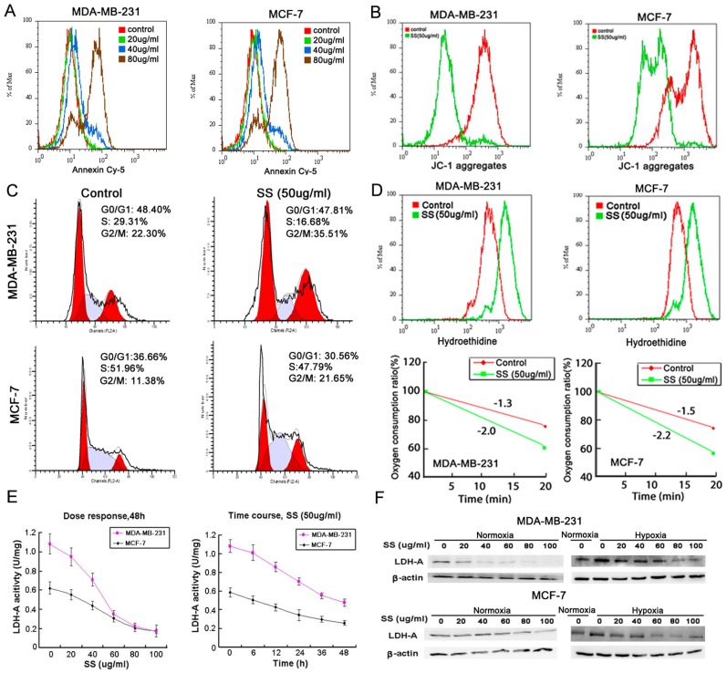
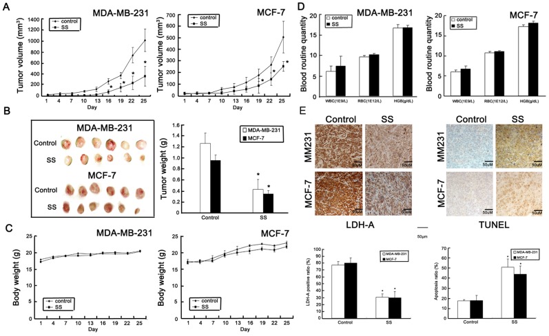
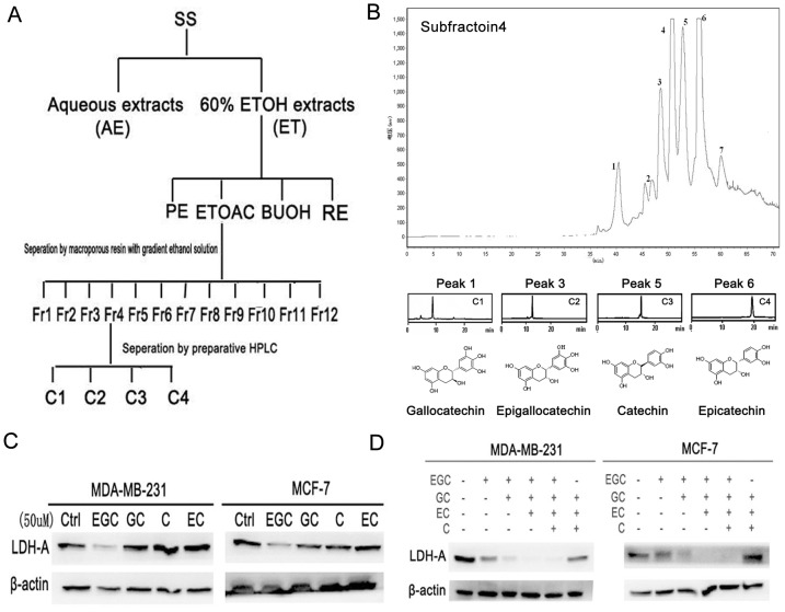
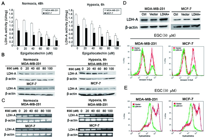
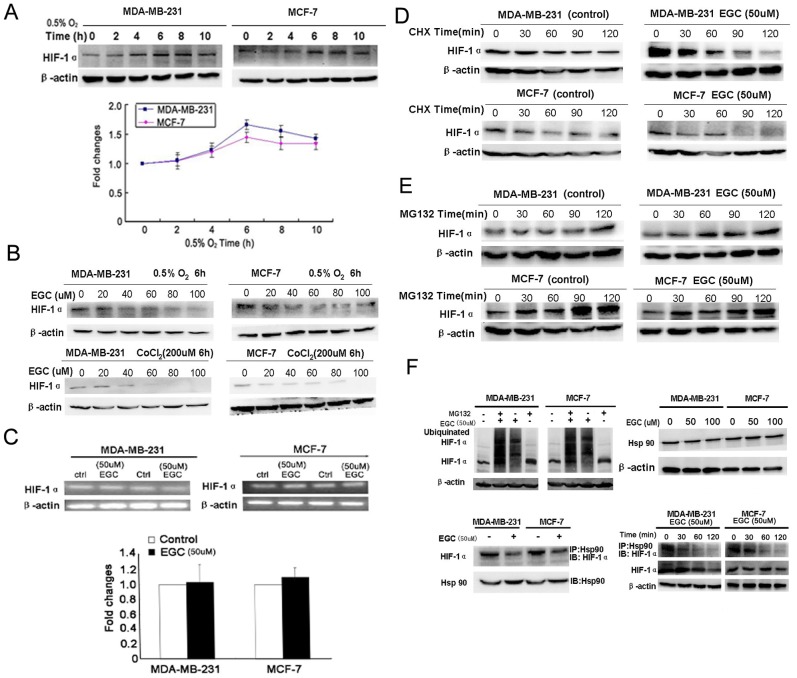
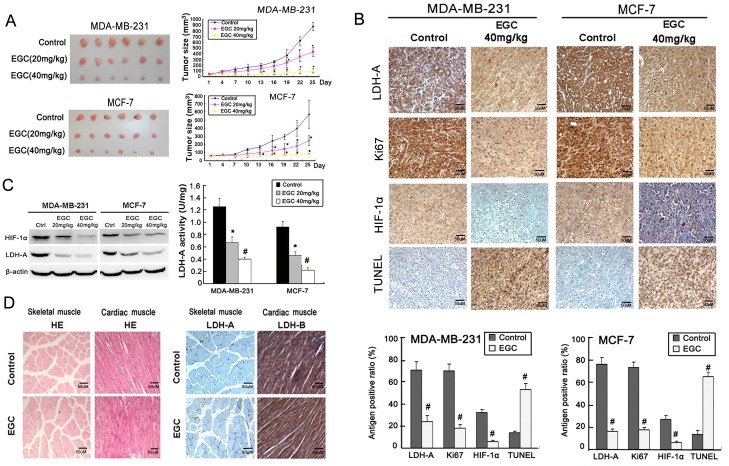
Aerobic glycolysis is an important feature of cancer cells. In recent years, lactate dehydrogenase A (LDH-A) is emerging as a novel therapeutic target for cancer treatment. Seeking LDH-A inhibitors from natural resources has been paid much attention for drug discovery. Spatholobus suberectus (SS) is a common herbal medicine used in China for treating blood-stasis related diseases such as cancer. This study aims to explore the potential medicinal application of SS for LDH-A inhibition on breast cancer and to determine its bioactive compounds. We found that SS manifested apoptosis-inducing, cell cycle arresting and anti-LDH-A activities in both estrogen-dependent human MCF-7 cells and estrogen-independent MDA-MB-231 cell. Oral herbal extracts (1 g/kg/d) administration attenuated tumor growth and LDH-A expression in both breast cancer xenografts. Bioactivity-guided fractionation finally identified epigallocatechin as a key compound in SS inhibiting LDH-A activity. Further studies revealed that LDH-A plays a critical role in mediating the apoptosis-induction effects of epigallocatechin. The inhibited LDH-A activities by epigallocatechin is attributed to disassociation of Hsp90 from HIF-1α and subsequent accelerated HIF-1α proteasome degradation. In vivo study also demonstrated that epigallocatechin could significantly inhibit breast cancer growth, HIF-1α/LDH-A expression and trigger apoptosis without bringing toxic effects. The preclinical study thus suggests that the potential medicinal application of SS for inhibiting cancer LDH-A activity and the possibility to consider epigallocatechin as a lead compound to develop LDH-A inhibitors. Future studies of SS for chemoprevention or chemosensitization against breast cancer are thus warranted.


Conflict of interest statement

Competing Interests: The authors have declared that no competing interests exist.

Figures

Figure 1
Figure 1. SS induces breast cancer cell apoptosis, G2/M checkpoint arrest, ROS accumulation and LDH-A inhibition.
(A) Annexin-Cy5 staining results showed that SS induced both breast cancer cells apoptosis in a dose-dependent manner; (B) JC-1 staining assay indicated that mitochondrial membrane potential was decreased after SS treatment, indicating that the mitochondrial pathway apoptosis was triggered by SS; (C) Cell cycle analysis showed that after SS treatment, the G2/M checkpoint was arrested in both breast cancer cell lines, presenting as a significant increase in G2/M subpopulations; (D) Hydroethidine was applied to detect the intracellular O2 level after SS administration by flow cytometry. The results showed that the intracellular O2 level was increased after SS treatment for 24 h (upper panel). Clark oxygen electrode was applied to detect the oxygen consumption speed of breast cancer cells after SS treatment. The results showed that the oxygen consumption speed of both breast cancer cells was rapidly enhanced after SS administration (lower panel); (E) LDH-A activity assay showed that SS were dose- and time- dependent when suppressing the LDH-A activity; (F) Western blotting results indicated that after SS administration, the expression of LDH-A in both breast cancer cells under both normoxia and hypoxia condition were down-regulated.
Figure 2
Figure 2. SS inhibits breast cancer growth in vivo.
(A) Tumor growth curve of both cancer xenografts during therapy period. The results indicated that SS (1 g/kg/d, oral intake) could significantly inhibit tumor growth in both ER negative and positive breast cancers; (B) Representative tumor pictures in control and SS treated groups. The average of tumor weight in SS treated group was significantly decreased in comparison to that in the control group; (C) Body weight fluctuation influenced by SS. SS-treated group had little influences on body weight compared to the control groups; (D) Blood routine quantity assay revealed that SS has little blood toxicity effects on mice; (E) Immunohisotchemistry method was applied to detect the LDH-A expression in both control and SS treated groups. The results indicated that LDH-A expression in both SS-treated breast cancer xenografts were significantly reduced, while TUNEL analysis indicated that the apoptosis ratio in SS –treated tumor samples was significantly increased. (×100, scale bar = 50 um; All values represents as Mean±SD, n = 6, *P<0.05 vs. control).
Figure 3
Figure 3. Bioactivity-guided fractionation of SS targeting on LDH-A.
(A) Isolation scheme of SS. Three parameters including LDH-A activity, LDH-A expression and apoptosis were selected for bioactive compounds screening; Crude 60% ethanol extracts of SS were firstly subjected to different polar solvents to yield PE, EtOAc, BuOH and RE fractions. After the first round screening, the EtOAc fraction was further separated with macroporous resin to yield 12 subfractions. Following the second round screening, subfraction 4 was then subjected to preparative HPLC and four compounds were finally purified. (B) Preparative HPLC chromatography of subfraction 4. A total of 7 single peaks were identified and the peaks 1, 3, 5 and 6 were finally purified through preparative HPLC. After Mass and NMR identification, the four compounds were identified as gallocatechin, epigallocatechin, catechin and epicatechin; (C) Among the four compounds, EGC exhibited the highest activity in inhibiting LDH-A expression; (D) EGC played synergistic role with the other three compounds in inhibiting LDH-A expression. The four compounds were added sequentially according to their relative concentrations in SS (10 µM ∶ 25 µM ∶ 200 µM ∶ 200 µM). When the four compounds were put together, they showed the highest inhibition effects on LDH-A expression. However, when EGC was withdrawn, the inhibition effects significantly weakened, indicating that EGC might play a key role in inhibiting LDH-A activity.
Figure 4
Figure 4. EGC inhibits breast cancer LDH-A activity and expression.
(A) LDH-A activity assay was applied to detect the effects of EGC on LDH-A activity in both breast cancer cell lines. The results indicated that EGC could dose- and time-dependently suppressed the LDH-A activity (All values represents as Mean± SD, n = 3, *P<0.05 vs. control); (B) The expression of LDH-A was also validated by Western blotting after EGC administration. The results showed that SS could inhibit LDH-A expression under both normoxia and hypoxia condition; (C) RT-PCR method was utilized to detect the level changes of LDH-A mRNA after EGC treatment. The results revealed that LDH-A mRNA levels were also down-regulated with increasing dose of EGC under both normoxia and hypoxia conditions; (D) The full length cDNA of LDH-A were amplified and subcloned into the pcDNA 3.1(+) vector. The over-expression of LDH-A was validated by Western blotting (upper panel). Annexin V-Cy5 staining analysis demonstrated that the EGC- induced apoptosis was reduced in LDH-A over-expression breast cancer cells; (E) Hydroethidine staining assay also revealed that in LDH-A over-expression breast cancer cells, the EGC-induced ROS elevation was also significantly eliminated.
Figure 5
Figure 5. EGC promotes HIF-1α proteasome pathway degradation.
(A) Expression curve of HIF-1α under hypoxia. The results showed that the expression level of HIF-1α reached the peak at the 6th hour, but had a slight decrease after 10 hours; (B) EGC inhibited HIF-1α expression in a dose-dependent manner in both breast cancer cells under both hypoxia condition and hypoxia mimic created by cobalt chloride; (C) Real-time PCR analysis was applied to detect the influences of EGC on HIF-1α mRNA levels. The results showed that there were little changes on HIF-1α mRNA level after EGC administration; (D) Cychloheximide (CHX) was administrated on breast cancer cells to inhibit protein synthesis. The expression of HIF-1α in both control and EGC treated cancer cells were validated by western blotting. The results showed that the HIF-1α degradation speed was much faster in EGC treated groups than in control groups, indicating that EGC might promote HIF-1α degradation; (E) MG132 was administrated on breast cancer cells to block the proteasome activity. The levels of HIF-1α were determined after MG132 treatment in both control and EGC treated groups. The results indicated that the accumulation quantities of HIF-1α were similar in both control and EGC treated groups, indicating that proteasome degradation was the main pathway accounting for decreased HIF-1α expression; (F) The ubiquinated HIF-1α level in both control and EGC treated group were detected by Western blotting. The results showed that EGC increased the expression level of ubiquinated HIF-1α in both cancer cells, indicating that EGC promoted the HIF-1α proteasome degradation pathway; Meanwhile, the interaction between HIF-1α and Hsp90 were validated by immunoblotting and immunoprecipitation assay. The results indicated that EGC brought little influences on Hsp90 expression. In EGC treated cell samples, the levels of HIF-1α binding to Hsp90 were decreased in comparison to control groups. Meanwhile, the dissociation of HIF-1α from Hsp90 was demonstrated to be an early event happed before downrgulation of HIF-1α induced by EGC, implying that EGC promotes HIF-1α degradation via interfering with the interaction between Hsp90 and HIF-1α. (All values represented as Mean± SD, n = 3, *P<0.05 vs. control).
Figure 6
Figure 6. EGC inhibits breast cancer growth and LDH-A expression in vivo.
(A) In MDA-MB-231 and MCF-7 breast cancer xenografts, EGC (20 mg/kg/d and 40 mg/kg/d) dose-dependently inhibited breast cancer growth, but had little influences on mice body weight; (B) The expression of LDH-A, Ki67 and HIF-1α in both control and EGC treated tumor samples were detected by immunohistochemistry. The results showed that in both breast cancer xenografts, the expression levels of LDH-A, Ki67 and HIF-1α were decreased in comparison to that in control groups, while apoptosis ratio was elevated in EGC-treated tumor samples detected by TUNEL method; (C) Western blotting analysis showed that the expression of HIF-1α and LDH-A in EGC-treated tumor samples were significantly decreased in comparison with control groups (left panel). The LDH-A activity in control and EGC-treated tumor samples were determined. The results showed that there was a significant reduction in LDH-A activity in EGC-treated tumor samples (right panel); (D) HE staining and immunohistochemistry assay indicated that EGC had little influences on the morphology and LDH expression in muscle and heart (×100, scale bar = 50 um; All values represents as Mean± SD, n = 6, *P<0.05 vs. control).

References

    1. Hanahan D, Weinberg RA (2011) Hallmarks of cancer: the next generation. Cell 144: 646–674. - PubMed
    1. Vander Heiden MG (2011) Targeting cancer metabolism: a therapeutic window opens. Nat Rev Drug Discov 10: 671–684. - PubMed
    1. Cairns RA, Harris IS, Mak TW (2011) Regulation of cancer cell metabolism. Nat Rev Cancer 11: 85–95. - PubMed
    1. Warburg O (1956) On the origin of cancer cells. Science 123: 309–314. - PubMed
    1. Pelicano H, Martin DS, Xu RH, Huang P (2006) Glycolysis inhibition for anticancer treatment. Oncogene 25: 4633–4646. - PubMed

Publication types

MeSH terms