. 2013;8(2):e56819.
doi: 10.1371/journal.pone.0056819. Epub 2013 Feb 14.

Inhibitory effects of resveratrol on PDGF-BB-induced retinal pigment epithelial cell migration via PDGFRβ, PI3K/Akt and MAPK pathways

Affiliations

Inhibitory effects of resveratrol on PDGF-BB-induced retinal pigment epithelial cell migration via PDGFRβ, PI3K/Akt and MAPK pathways

Chi-Ming Chan et al. PLoS One. 2013.

Abstract

Purpose: In diseases such as proliferative vitreoretinopathy (PVR), proliferative diabetic retinopathy, and age-related macular degeneration, retinal pigment epithelial (RPE) cells proliferate and migrate. Moreover, platelet-derived growth factor (PDGF) has been shown to enhance proliferation and migration of RPE cells in PVR. Even resveratrol can suppress the migration and adhesion of many cell types, its effects on RPE cell migration and adhesion remain unknown. In this study, we investigated the inhibitory effects of resveratrol on RPE cell migration induced by PDGF-BB, an isoform of PDGF, and adhesion to fibronectin, a major ECM component of PVR tissue.

Methods: The migration of RPE cells was assessed by an electric cell-substrate impedance sensing migration assay and a Transwell migration assay. A cell viability assay was used to determine the viability of resveratrol treated-cells. The cell adhesion to fibronectin was examined by an adhesion assay. The interactions of resveratrol with PDGF-BB were analyzed by a dot binding assay. The PDGF-BB-induced signaling pathways were determined by western blotting and scratch wound healing assay.

Results: Resveratrol inhibited PDGF-BB-induced RPE cell migration in a dose-dependent manner, but showed no effects on ARPE19 cell adhesion to fibronectin. The cell viability assay showed no cytotoxicity of resveratrol on RPE cells and the dot binding assay revealed no direct interactions of resveratrol with PDGF-BB. Inhibitory effects of resveratrol on PDGF-BB-induced platelet-derived growth factor receptor β (PDGFRβ) and tyrosine phosphorylation and the underlying pathways of PI3K/Akt, ERK and p38 activation were found; however, resveratrol and PDGF-BB showed no effects on PDGFRα and JNK activation. Scratch wound healing assay demonstrated resveratrol and the specific inhibitors of PDGFR, PI3K, MEK or p38 suppressed PDGF-BB-induced cell migration.

Conclusions: These results indicate that resveratrol is an effective inhibitor of PDGF-BB-induced RPE cell migration via PDGFRβ, PI3K/Akt and MAPK pathways, but has no effects on the RPE cell adhesion to fibronectin.


Conflict of interest statement

Competing Interests: The authors have declared that no competing interests exist.

Figures

Figure 1
Figure 1. PDGF-BB-induced ARPE19 cell migration was inhibited by resveratrol in an ECIS migration assay.
The cells cultured in 8W1E ECIS arrays were treated with different combinations of PDGF-BB (20 ng/ml) and resveratrol (CTL indicates that it contained only DMSO) which were preincubated together at 37°C for 30 minutes. Cell migration was then assessed by continuous resistance measurements for 30 hours. Resveratrol (Res) (10 µM) did not increased cell migration when PDGF-BB was not present. In the well containing PDGF-BB but not resveratrol, the impedance, which corresponds to the number of cells migrated to the surface of the detective electrode, increased sharply during the first 10 hours. By contrast, the impedance in the well containing PDGF-BB and resveratrol increased slowly during the same time period.
Figure 2
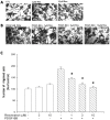
Figure 2. Transwell migration assay showed that PDGF-BB-induced ARPE19 cell migration was inhibited by resveratrol.
Transwell inserts were coated with fibronectin (0.3 mg). ARPE19 cells (5×10⁴ in 200 µl) were seeded in the upper chamber in the absence or presence of resveratrol. The inserts were assembled in the lower chamber, which was filled with 600 µl serum-free medium without PDGF-BB (A) and containing PDGF-BB (20 ng/ml) (B), and preincubated with various concentrations of resveratrol for 30 mininutes at 37°C. After incubating for 5 hours at 37°C, fixation was performed. ARPE19 cells that migrated to the underside of filter membrane were photographed (A, B) and counted by phase contrast light microscope under high power field (magnification, 100×), (C). All experiments were conducted in duplicates and similar results were repeated four times. The results are expressed as percentage of control and represent mean ± standard errors (SE) of the eight experiments. *p<0.05 significantly differs from PDGF-BB-stimulated cells (the fourth bar).
Figure 3
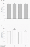
Figure 3. Viability and cell adhesion of ARPE19 cells was not influenced by resveratrol.
The cells were treated with different concentrations of resveratrol for 24 hours after being starved for 24 hours. Cell viability was determined by MTT assay (A). BCECF-labeled cells were treated with DMSO or resveratrol for 30 minutes. They were then seeded and allowed to adhere on plates with precoated fibronectin (fn) (15 µg/ml) at 37°C for 1 hour. Fluorescence was measured using excitation and emission wavelength of 485 and 535 nm, respectively (B). The results are expressed as percentage of control and represent the mean ± standard errors (SE) of four independent experiments.
Figure 4
Figure 4. Resveratrol did not directly interact with PDGF-BB in dot binding assay.
Human recombinant PDGF-BB, phosphate buffer saline (PBS) and the indicated concentrations of resveratrol were applied onto the nitrocellulose (NC) membrane. The membrane was incubated with PDGF-BB in PBS and then developed by probing with Ab directed against PDGF-BB. The results presented are representative of four independent experiments.
Figure 5
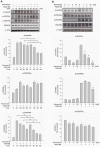
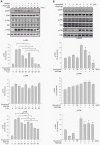
Figure 5. PDGF-BB-induced tyrosine and PDGFRβ phosphorylations were inhibited by resveratrol in a time- and concentration-dependant manner.
ARPE19 cells were treated with the indicated lengths of time of PDGF-BB (20 ng/ml) and preincubated with or without resveratrol (10 µM) at 37°C (A). After being further preincubated for the indicated concentrations of resveratrol and incubated with or without PDGF-BB (20 ng/ml) at 37°C for 30 minutes, the cells were collected and their lysates were analyzed by Western blot analysis (B). The changes in phosphorylated tyrosine, PDGFRα and PDGFRβ expression were evaluated. The quantitative data of western blot are shown below the panels which are expressed as percentage of control and represent mean ± standard errors (SE) of the four independent experiments. *p<0.05 significantly differs from same indicated time of cells stimulated PDGF-BB only (A) and *p<0.05 significantly differs from PDGF-BB-stimulated cells (the fifth bar) (B).
Figure 6
Figure 6. PDGF-BB-induced PI3K and Akt phosphorylations were inhibited by resveratrol in a time- and concentration-dependant manner.
ARPE19 cells were treated with the indicated lengths of time of PDGF-BB (20 ng/ml) and preincubated with or without resveratrol (10 µM) at 37°C (A). After being further preincubated for the indicated concentrations of resveratrol and incubated with or without PDGF-BB (20 ng/ml) at 37°C for 30 minutes, the cells were collected and their lysates were analyzed by Western blot analysis (B). The changes in phosphorylated PI3K and Akt expression were evaluated. The quantitative data of western blot are shown below the panels which are expressed as percentage of control and represent mean ± standard errors (SE) of the four independent experiments. *p<0.05 significantly differs from same indicated time of cells stimulated PDGF-BB only (A) and *p<0.05 significantly differs from PDGF-BB-stimulated cells (the fifth bar) (B).
Figure 7
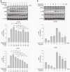
Figure 7. PDGF-BB-induced ERK and P38 phosphorylations were inhibited by resveratrol in a time- and concentration-dependant manner.
ARPE19 cells were treated with the indicated lengths of time of PDGF-BB (20 ng/ml) and preincubated with or without resveratrol (10 µM) at 37°C (A). After being further preincubated for the indicated concentrations of resveratrol and incubated with or without PDGF-BB (20 ng/ml) at 37°C for 30 minutes, the cells were collected and their lysates were analyzed by Western blot analysis (B). The changes in phosphorylated ERK, JNK and p38 expression were evaluated. The quantitative data of western blot are shown below the panels which are expressed as percentage of control and represent mean ± standard errors (SE) of the four independent experiments. *p<0.05 significantly differs from same indicated time of cells stimulated PDGF-BB only (A) and *p<0.05 significantly differs from PDGF-BB-stimulated cells (the fifth bar) (B).
Figure 8
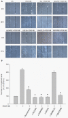
Figure 8. PDGF-BB-induced cell migrations were inhibited by resveratrol and by suppression of PDGFR, PI3K/Akt and MAPK signaling.
The plates with confluent monolayer of ARPE cells were pretreated with mitomycin-C (5 µg/ml) for 1 hour, then wounded with a linear scratching by a sterile 20- µl pipette tip. The cells were immediately washed and were incubated with PDGF-BB (20 ng/ml) only, PDGF-BB in combination with resveratrol (10 µM), AG1295 (10 µM), LY294002 (10 µM), U0126 (10 µM), SP600125 (3 µM) and SB203580 (3 µM) respectively. The wound closure was monitored for 16 h followed by photography under phase-contrast microscope (x100) (A). The quantitative data of the number of migrated cell in the wound area are expressed as percentage of control and represent mean ± standard errors (SE) of the four independent experiments. *p<0.05 significantly differs from PDGF-BB-stimulated cells (the second bar) (B).

References

    1. Ho J, Witkin AJ, Liu J, Chen Y, Fujimoto JG, et al. (2011) Documentation of intraretinal retinal pigment epithelium migration via high-speed ultrahigh-resolution optical coherence tomography. Ophthalmology 118: 687–693. - PMC - PubMed
    1. Charteris DG, Sethi CS, Lewis GP, Fisher SK (2002) Proliferative vitreoretinopathy-developments in adjunctive treatment and retinal pathology. Eye (Lond) 16: 369–374. - PubMed
    1. Cardillo JA, Stout JT, LaBree L, Azen SP, Omphroy L, et al. (1997) Post-traumatic proliferative vitreoretinopathy. The epidemiologic profile, onset, risk factors, and visual outcome. Ophthalmology 104: 1166–1173. - PubMed
    1. Campochiaro PA (1997) Pathogenic mechanisms in proliferative vitreoretinopathy. Arch Ophthalmol 115: 237–241. - PubMed
    1. de Silva DJ, Kwan A, Bunce C, Bainbridge J (2008) Predicting visual outcome following retinectomy for retinal detachment. Br J Ophthalmol 92: 954–958. - PubMed

Publication types

MeSH terms