Review
. 2014 Jan;224(1):29-35.
doi: 10.1111/joa.12034. Epub 2013 Mar 4.

The role of muscle-specific tyrosine kinase (MuSK) and mystery of MuSK myasthenia gravis

Affiliations
Review

The role of muscle-specific tyrosine kinase (MuSK) and mystery of MuSK myasthenia gravis

Inga Koneczny et al. J Anat. 2014 Jan.

Abstract

MuSK myasthenia gravis is a rare, severe autoimmune disease of the neuromuscular junction, only identified in 2001, with unclear pathogenic mechanisms. In this review we describe the clinical aspects that distinguish MuSK MG from AChR MG, review what is known about the role of MuSK in the development and function of the neuromuscular junction, and discuss the data that address how the antibodies to MuSK lead to neuromuscular transmission failure.

Keywords: AChR; DOK7; IgG4; LRP4; MG; RAPSN; muscle-specific tyrosine kinase; myasthenia gravis; neuromuscular transmission; quantal content.


Figures

Fig. 1
Fig. 1
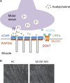
(A) The neuromuscular junction illustrating the ACh located in synaptic vesicles and the high density of acetylcholine receptors (AChRs) clustered by RAPSN on the tops of the folds of postsynaptic membrane (grey). The AGRN/LRP4/MuSK/DOK7 pathway is essential for the clustering process. (B) The pathway and the action of MuSK antibodies can be studied in vitro by applying AGRN to C2C12 myotubes. In the presence of medium or healthy control serum samples (HC), AGRN induces clusters of AChRs, identified with fluorescent bungarotoxin. In the presence of MuSK antibodies, there are very few clusters formed.
Fig. 2
Fig. 2
Anterograde and retrograde signalling at the neuromuscular junction. AGRN is released by the nerve and binds to LRP4, which then binds to MuSK. This interaction leads to MuSK autophosphorylation and activation of its kinase function, leading to anterograde signalling by subsequent phosphorylation of DOK7 (not shown), which binds MuSK as a dimer. RAPSN clusters the AChR adjacent to the nerve terminal. LRP4 is thought to send a retrograde signal for development of the presynaptic site, including clustering voltage-gated calcium channels (VGCC) and other presynaptic molecules such as Bassoon and Piccolo and vesicles containing ACh at the nerve terminal.

References

    1. Arber S, Burden SJ, Harris AJ. Patterning of skeletal muscle. Curr Opin Neurobiol. 2002;12:100–103. - PubMed
    1. ter Beek WP, Martinez-Martinez P, Losen M, et al. The effect of plasma from muscle-specific tyrosine kinase myasthenia patients on regenerating endplates. Am J Pathol. 2009;175:1536–1544. - PMC - PubMed
    1. Bergamin E, Hallock PT, Burden SJ, et al. The cytoplasmic adaptor protein Dok7 activates the receptor tyrosine kinase MuSK via dimerization. Mol Cell. 2010;39:100–109. - PMC - PubMed
    1. Burden SJ. Building the vertebrate neuromuscular synapse. J Neurobiol. 2002;53:501–511. - PubMed
    1. Burden SJ. SnapShot: neuromuscular junction. Cell. 2011;144:826–826.e1. - PubMed

Publication types

MeSH terms

LinkOut - more resources