Review
. 2013 Dec;34(6):1088-108.
doi: 10.1016/j.mam.2013.02.002. Epub 2013 Feb 28.

Structural biology of the writers, readers, and erasers in mono- and poly(ADP-ribose) mediated signaling

Affiliations
Review

Structural biology of the writers, readers, and erasers in mono- and poly(ADP-ribose) mediated signaling

Tobias Karlberg et al. Mol Aspects Med. 2013 Dec.

Abstract

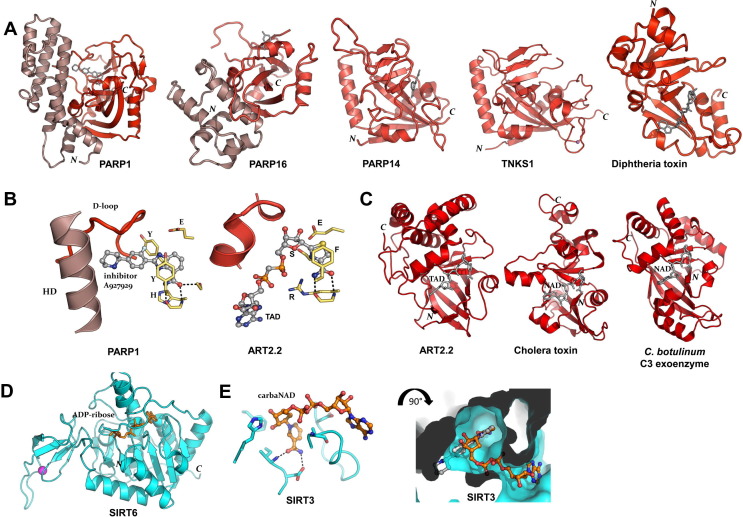
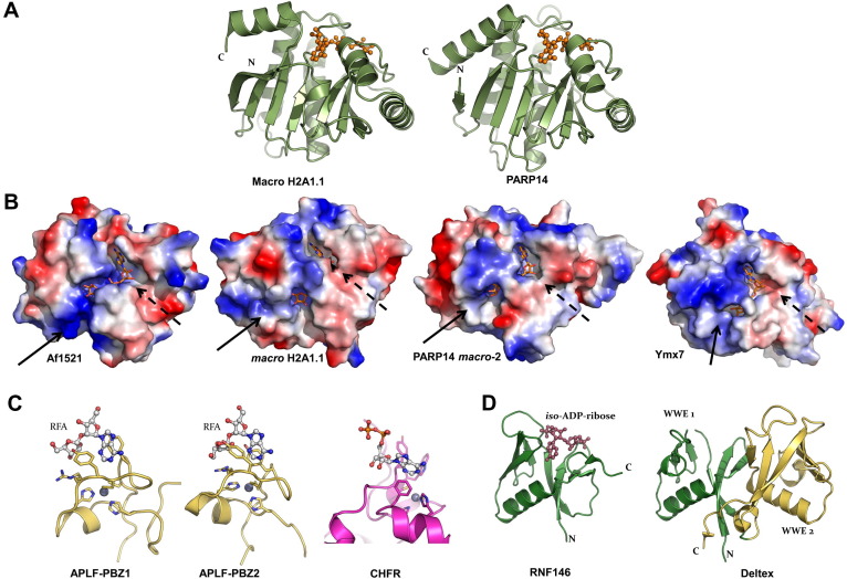
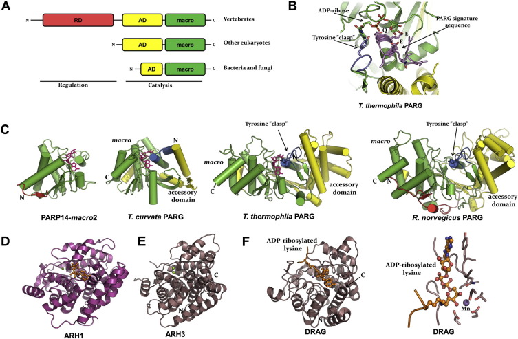
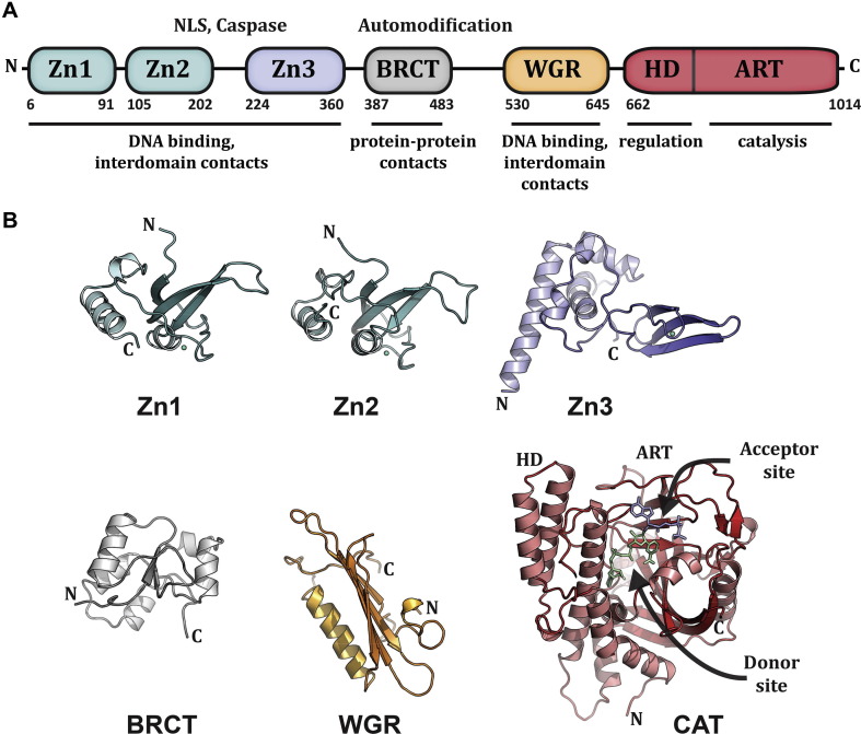
ADP-ribosylation of proteins regulates protein activities in various processes including transcription control, chromatin organization, organelle assembly, protein degradation, and DNA repair. Modulating the proteins involved in the metabolism of ADP-ribosylation can have therapeutic benefits in various disease states. Protein crystal structures can help understand the biological functions, facilitate detailed analysis of single residues, as well as provide a basis for development of small molecule effectors. Here we review recent advances in our understanding of the structural biology of the writers, readers, and erasers of ADP-ribosylation.

Keywords: ADP-ribosylation; DNA repair; Glycohydrolase; Macro domain; Signaling pathways; Transferase.


Figures

Fig. 1
Fig. 1
Structures of ADP-ribosyl transferases. (A) The ARTD family. Crystal structures of the transferase domains of human PARP1 (PDB: 3L3M), PARP16 (PDB: 4F0D), PARP14 (PDB: 4F1L), TNKS1 (PDB: 2RF5), all with nicotinamide mimicking inhibitors, and diphtheria toxin in complex with NAD+ (PDB: 1TOX). Two central β-sheets, one five stranded anti-parallel and one four stranded mixed β-sheet make up the core of the transferase domain. The β-sheets are surrounded by α-helices on each side and both modules contribute to the NAD+ binding crevice. An additional helical domain in PARP1 and PARP16 are shown in darker shade. N- and C-terminal positions are indicated. (B) Structure surrounding the signature motifs that contribute to the active site. Left, the H-Y-Y/F-E motif exemplified by ARTD member PARP1 in complex with inhibitor A927929 (PDB: 3L3M), with part of the α-helical domain indicated; right, the R-S-F-E motif exemplified by ARTC member rat ART2.2 in complex with NAD+-analog TAD+ (PDB: 1OG1). (C) The ARTC family. Crystal structures of the transferase domains of rat ecto-ART2.2 in complex with the NAD+-analog TAD+ (PDB: 1OG1), cholera toxin A1 in complex with NAD+ (PDB: 2A5F) and Clostridium botulinum C3 exoenzyme in complex with NAD+ (PDB: 2A9K). (D) The sirtuin family: human SIRT6 in complex with ADP-ribose (PDB: 3PKI). (E) Sirtuin NAD+ binding mode illustrated by human SIRT3 in complex with carba-NAD+ (PDB: 4FVT). Note the absence of nicotinamide stacking aromatic sidechains as compared to panel B. Right, the same structure, surface rendered and rotated to view into the nicotinamide pocket. Superimposition of the PARP inhibitor from panel B (in grey) illustrate that sirtuin ligands bind in a conformation different from that of ARTD and ARTC ligands.
Fig. 2
Fig. 2
Structures of ADP-ribosyl binder domains. (A) Crystal structures of the macro domain of human histone macro H2A1.1 (PDB: 3IID) and of human PARP14 macro domain 2 (PDB: 3Q71), both in complex with ADP-ribose. (B) Charge potential surface rendering of Af1521 (PDB: 2BFQ), histone macro H2A1.1, PARP14 macro domain 2, and yeast Ymx7 (PDB: 1TXZ), all in complex with ADP-ribose. Solid arrows indicate the putative PAR crevice or its corresponding position; dashed arrows indicate the either exposed or buried adenine ribose site. (C) Structures of the two PBZ domains of APLF (PDB: 2KQD, 2KQE) in complex with ribofuranosyladenosine (RFA) and the PBZ domain of CHFR (PDB: 2XOC). (D) Crystal structures of the WWE domain of E3 ubiqutin protein ligase RNF146 in complex with iso-ADP-ribose (PDB: 3V3L) and the tandem WWE-domain of Drosophila melanogaster Deltex protein (PDB: 2A90).
Fig. 3
Fig. 3
Structures of ADP-ribosyl erasers. (A) Schematic domain arrangement in PARG orthologs. RD, regulary domain; AD, accessory domain; macro, macro homology domain. (B) Closeup of the ADP-ribose binding site including the PARG signature motif in a eukaryotic PARG (T. thermophila; PDB: 4EPP). Green and yellow indicate domains as in panel A. (C) Crystal structures of PARP14 macro domain 2 (PDB: 3Q71), a bacterial PARG (PDB: 3SIJ), and the T. thermophila and rat orthologs (PDB: 3UEK). Colors indicate domains as in panel A. (D) Structure of human ARH1 (PDB: 3HFW). (E) Structure of human ARH3 (PDB: 2FOZ). (F) Structure of the dinitrogenase reductase activating glycohydrolase (DRAG) from Rhodospirillum rubrum in complex with ADP-ribosyllysine (PDB: 2WOD), and right, closeup view of ADP-ribosyllysine trapped in the active site of DRAG.(For interpretation of the references to colour in this figure legend, the reader is referred to the web version of this book.)
Fig. 4
Fig. 4
PARP1 modular domain structure. (A). Schematic representation of human PARP1 domains. The amino acid numbering for human domain boundaries are noted. Homologous DNA-binding zinc finger domains, Zn1 and Zn2, are located at the N-terminus of PARP1. These domains are followed by a linker region containing a bipartite nuclear localization signal (NLS) and a caspase three cleavage site (Caspase). This regulatory region is followed by a third zinc finger domain (Zn3), which has a distinct structure and function from that of Zn1 and Zn2. A BRCA C-terminus (BRCT) fold is located within the region of PARP1 that is primarily targeted for automodification. The C-terminal end of PARP1 contains a WGR domain, named after a conserved Trp-Gly-Arg sequence, and the catalytic domain, which is composed of an α-helical subdomain (HD) and an ADP-ribosyl transferase subdomain (ART). The known functions for the individual domains are noted beneath the schematic. (B). Crystal and/or NMR structures have been determined for each of the PARP1 domains in the absence of DNA: the NMR structures for the homologous Zn1 and Zn2 domains [PDB: 2DMJ and 2CS2; see also reference (Eustermann et al., 2011)], the NMR structure of the Zn3 domain is shown [PDB: 2JVN; see also (Langelier et al., 2008) for crystal structure determination], the NMR structure of the BRCT fold [PDB: 2COK; see also (Loeffler et al., 2011), the NMR structure of the WGR domain (PDB: 2CR9), and the crystal structure of the catalytic domain (PDB: 1A26) (Ruf et al., 1996)]. Catalytic subdomains are labeled (HD and ART). The crystal structure was determined in the presence of an NAD+ analog that has defined the proposed “acceptor site” for poly(ADP-ribose) formation. The NAD+ binding site is modeled in this figure based on the structure of an ADP-ribosylating toxin structure (Bell and Eisenberg 1996). Reprinted from (Langelier and Pascal 2013) with permission from Elsevier.
Fig. 5
Fig. 5
Structures of PARP1 domains bound to DNA damage. (A) Crystal structure of human Zn1 domain in complex with a DNA double strand break (PDB: 3OD8). (B) Crystal structure of human Zn2 domain in complex with a DNA double strand break (PDB: 3ODC). Together, the structures in panels A and B illustrate the features of damage DNA that are recognized by PARP1 zinc finger domains: a continuous phosphate backbone engaged primarily by conserved Arg (R) residues on the backbone grip, and exposed nucleotide bases engaged by hydrophobic residues on the base stacking loop. Key Zn1 residues that are involved in communicating with other PARP1 domains are shown (D45 and W79). (C) The Zn1, Zn3, and WGR domains collectively assemble on a DNA double strand break, with each domain forming specific protein-DNA, and protein–protein contacts with adjacent domains. The Zn1 domain has the same orientation as shown in panel A, and the same key residues are shown as sticks. WGR residues W589 and R591 are essential to PARP1 DNA-dependent activity and form protein-DNA and protein–protein contacts, respectively. Zn1 residue W79 is critical for PARP1 DNA-dependent activity and is located at the interface with Zn3. (D) Crystal structure of the Zn1–Zn2 domains of PARP1 bound to a DNA double strand break with a single 5′ nucleotide overhang. In this complex, the Zn2 domain forms contacts with the DNA similar to that seen for the isolated domain in panel B (the same conserved residues are shown). The Zn1 domain binds to DNA with a opposite polarity to that seen in panels A and C, with Arg18 inserting into the major groove rather than the minor groove, and the base stacking loop forming protein–protein contacts rather than protein-DNA contacts. Reprinted from (Langelier and Pascal 2013) with permission from Elsevier.
Fig. 6
Fig. 6
Crystal structure of essential PARP1 domains in complex with DNA damage. (A) Human PARP1 domains Zn1, Zn3, and WGR-CAT were crystallized in complex with a DNA double-strand break (PDB: 4DQY) (Langelier et al., 2012). The PARP1/DNA complex illustrates how DNA damage detection is coupled to structural transitions in the catalytic domain that elevate poly(ADP-ribosyl)ation activity (Langelier et al., 2012). Three interdomain contact regions form upon PARP1 interaction with DNA. The Zn1-WGR-HD interface is the most direct coupling of DNA damage recognition to the catalytic domain, with Zn1 contacting both DNA damage and WGR, and the WGR contacting the HD. The Zn3-WGR-HD interface forms where the zinc ribbon motif of Zn3 contacts conserved WGR and HD residues, with W318 as a central component of the interaction. The Zn1-Zn3 interface forms where these two domains rest together on DNA. Mutations that target these interfaces have a severe impact on DNA damage-dependent PARP1 activity (Langelier et al., 2010, Langelier et al., 2011, Langelier et al., 2012), indicating that the structure has captured interdomain contacts that are relevant to PARP1 regulation. Collectively, the interdomain communication with the HD displaces conserved Leu residues from the hydrophobic interior of the HD, leading to destabilization of the CAT that correlates with an elevation in PARP1 catalytic activity. (B) A model for the approximate positioning of the Zn2 and BRCT domains within the PARP1/DNA complex. Zn1, Zn3, and WGR-CAT are shown as surfaces, labeled, and colored as in Fig. 4A. The Zn2 and BRCT domains are drawn in schematic representation. Their positioning is based on the relative location of the termini of adjacent domains in the structure. The numbering and location of linker residues are shown. The arrow indicates the location of the PARP1 automodification region near the catalytic active site. Reprinted from (Langelier and Pascal 2013) with permission from Elsevier.
Fig. 7
Fig. 7
Tankyrase domain structure. (A) Tankyrase 1 and 2 have a similar domain construction. The C-terminus contains a SAM domain implicated in tankyrase oligomerization and the PARP catalytic domain that synthesizes poly(ADP-ribose). The N-terminus is primarily composed of an ankyrin repeat region that is segmented into five ankyrin repeat clusters (ARC1 through ARC5), each containing five ankyrin modules (numbered 1–5 in ARC4). The ARCs interact with proteins targeted for modification by tankyrase, with the exception of ARC3 that lacks key conserved residues involved in target binding. The extreme N-terminus of tankyrase 1 contains runs of His, Pro, and Ser and is thus termed the HPS domain. HPS likely serves a regulatory function. Tankyrase 2 does not have an HPS domain. The residue numbering of domain boundaries is shown for tankyrase 1. (B) The crystal structure of ARC4 from tankyrase 2 illustrates the organization and specialization of the five ankyrin repeat modules that form each ARC (Guettler et al., 2011). Three typical ankyrin repeats form the core of the ARC (ankyrin modules 2–4). Modified ankyrin repeats initiate the N-terminus of the ARC (module 1, N-cap) and terminate the C-terminus of the ARC (module 5), thus capping each end of the ARC and preventing continuous stacking of ankyrin repeats. (C) X-ray structure of the catalytic domain of tankyrase 2 bound to inhibitor XAV939 (Karlberg et al., 2010b).
Fig. 8
Fig. 8
Structural basis for ARC recognition of target peptides. (A) The X-ray structure of ARC4 of tankyrase 2 in complex with a peptide derived from the target 3BP2 (Guettler et al., 2011), an adaptor protein in signaling pathways that depend on tankyrase activity for protein turnover. The core ankyrin repeats form the peptide interaction surface. An “arginine cradle” forms around the conserved Arg residue at position 1 of the consensus peptide sequence. Two Tyr residues form an “aromatic glycine sandwich” that surrounds the conserved Gly residue at position 6 of the consensus peptide sequence. (B) The amino acid sequence for 3BP2 is shown for each of the eight positions. Underlined residues are mutated in individuals with cherubism (Levaot et al., 2011), resulting in a loss of interaction with and modification by tankyrase. An optimized sequence was determined through an extensive analysis of amino acid substitutions at each of the eight positions.
Fig. 9
Fig. 9
Variations in ARC recognition of target peptides and organization of multiple ARCs. (A) The X-ray structure of the ARC2–ARC3 fragment of tankyrase 1 was determined in complex with the tankyrase binding region of Axin1, a key component of the Wnt signaling complex (Morrone et al., 2012). Two regions of Axin1, segment-N and segment-C, each interact with an ARC2 of tankyrase, thus forming a 1:2 complex of Axin1:tankyrase. An ARC2–ARC3 homodimer is formed where the ARC3N-cap of one monomer “crosses over” to stack against the core ankyrin modules of ARC3 in the second monomer. (B) The segment-N and segment-C interactions with ARC2 exhibit variations in how the polypeptides engage the target protein docking site. (C) Comparison of the amino acid sequences for segment-N and segment-C of Axin1. Segment-C has an insertion of 9 amino acids between position 1 and position 4, but otherwise has the same key features of the segment-N interaction with the ARC2, such as the arginine at position 1 and the glycine at position 6.

References

    1. Ahel D., Horejsi Z., Wiechens N., Polo S.E., Garcia-Wilson E., Ahel I., Flynn H., Skehel M., West S.C., Jackson S.P., Owen-Hughes T., Boulton S.J. Poly(ADP-ribose)-dependent regulation of DNA repair by the chromatin remodeling enzyme ALC1. Science. 2009;325:1240–1243. - PMC - PubMed
    1. Ahel I., Ahel D., Matsusaka T., Clark A.J., Pines J., Boulton S.J., West S.C. Poly(ADP-ribose)-binding zinc finger motifs in DNA repair/checkpoint proteins. Nature. 2008;451:81–85. - PubMed
    1. Ahuja N., Schwer B., Carobbio S., Waltregny D., North B.J., Castronovo V., Maechler P., Verdin E. Regulation of insulin secretion by SIRT4, a mitochondrial ADP-ribosyltransferase. J. Biol. Chem. 2007;282:33583–33592. - PubMed
    1. Ali A.A., Timinszky G., Arribas-Bosacoma R., Kozlowski M., Hassa P.O., Hassler M., Ladurner A.G., Pearl L.H., Oliver A.W. The zinc-finger domains of PARP1 cooperate to recognize DNA strand breaks. Nat. Struct. Mol. Biol. 2012;19:685–692. - PMC - PubMed
    1. Allen M.D., Buckle A.M., Cordell S.C., Lowe J., Bycroft M. The crystal structure of AF1521 a protein from Archaeoglobus fulgidus with homology to the non-histone domain of macroH2A. J. Mol. Biol. 2003;330:503–511. - PubMed

Publication types

MeSH terms

LinkOut - more resources