Review
. 2013 Apr;9(4):211-21.
doi: 10.1038/nrneurol.2013.29. Epub 2013 Mar 5.

Chronic neuropathologies of single and repetitive TBI: substrates of dementia?

Affiliations
Review

Chronic neuropathologies of single and repetitive TBI: substrates of dementia?

Douglas H Smith et al. Nat Rev Neurol. 2013 Apr.

Abstract

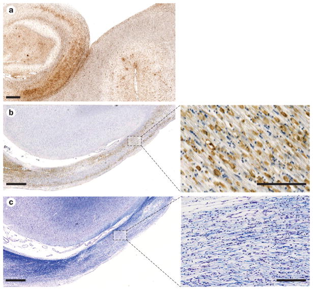
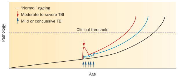
Traumatic brain injury (TBI) has long been recognized to be a risk factor for dementia. This association has, however, only recently gained widespread attention through the increased awareness of 'chronic traumatic encephalopathy' (CTE) in athletes exposed to repetitive head injury. Originally termed 'dementia pugilistica' and linked to a career in boxing, descriptions of the neuropathological features of CTE include brain atrophy, cavum septum pellucidum, and amyloid-β, tau and TDP-43 pathologies, many of which might contribute to clinical syndromes of cognitive impairment. Similar chronic pathologies are also commonly found years after just a single moderate to severe TBI. However, little consensus currently exists on specific features of these post-TBI syndromes that might permit their confident clinical and/or pathological diagnosis. Moreover, the mechanisms contributing to neurodegeneration following TBI largely remain unknown. Here, we review the current literature and controversies in the study of chronic neuropathological changes after TBI.


Conflict of interest statement

Competing interests

The authors declare no competing interests.

Figures

Figure 1
Figure 1
Cerebral atrophy following survival from a single moderate to severe TBI. a | Brain of a 24-year-old patient who died within hours of being assaulted. The only macroscopic evidence of TBI is a superficial contusion in the right inferior temporal lobe (arrow), although grade 1 diffuse traumatic axonal injury was observed on histology. b | Brain of a 40-year-old patient who survived 4 years from injury and has a healed contusion in a similar location to that in part a (arrow). In comparison to the previous patient, however, there is notable gyral atrophy, ventricular enlargement, and thinning of the corpus callosum. Abbreviation: TBI, traumatic brain injury.
Figure 2
Figure 2
Tau and amyloid-β pathology after TBI. a,b | Extensive neurofibrillary tangles observed in a boxer. The 77-year-old man had participated in over 700 boxing contests during his life and had neuropsychiatric symptoms. Tangles were identified using silver staining (a) and Congo red staining (b). c,d | Neurofibrillary tangles in the parahippocampal gyrus of a 47-year-old man who had sustained a single severe TBI 1 year previously. No history of repetitive TBI was recorded. Tangles were identified using immunohistochemistry (c) and thioflavine S staining (d). Scale bars 100 μm. e,f | Sections of the temporal cortex from a 63-year-old man with dementia pugilistica who had participated in over 300 boxing contests and was originally noted as plaque-negative. Extensive amyloid-β plaques were demonstrated using immunohistochemistry with formic acid pretreatment (e), but not with Congo red (f). Abbreviation: TBI, traumatic brain injury. Permission for parts a and b obtained from Cambridge University Press © Corsellis, J. A. et al. Psychol. Med. 3, 270–303 (1973). Permission for parts c and d obtained from John Wiley and Sons © Johnson, V. E. et al. Brain Pathol. 22, 142–149 (2012). Parts e and f reproduced from Roberts, G. W. et al. The occult aftermath of boxing. J. Neurol. Neurosurg. Psychiatry 53, 373–378 © 1990 with permission from BMJ Publishing Group Ltd.
Figure 3
Figure 3
Neuroinflammation and white matter degeneration after TBI. a | Extensive reactive microglia (immunostained with antibody CR3/43) in the hippocampus and adjacent sulci of a 65-year-old male with dementia pugilsitica. Scale bar 1 mm. b | Extensive CR3/43-reactive cells with an amoeboid morphology, indicative of macrophages, observed in the atrophic corpus callosum of a 37-year-old male 4 years following a single severe TBI. Scale bar 1 mm for main image (left) and 100 μm for high-magnification image (right). c | Adjacent section to b, stained with Luxol fast blue, indicating chronic white matter change and loss of myelin. Scale bar 1 mm for main image (left) and 100 μm for high-magnification image (right). Abbreviation: TBI, traumatic brain injury.
Figure 4
Figure 4
Interaction between TBI and ‘normal’ ageing: a hypothesis. An increasing range of pathologies is recognized in association with survival from TBI, any of which may contribute to the associated clinical syndromes of neurocognitive impairment. In the absence of a history of TBI, many of these same pathologies, such as amyloid-β plaque or neuroinflammatory pathology, may accumulate as a consequence of ’normal’ ageing (black line), with the accumulated pathologies eventually crossing a threshold where clinical symptoms are apparent. Following a single moderate to severe TBI, evidence to date supports an initial spike in pathology immediately after the event, which subsequently resolves. However, it is intriguing to speculate that, in a proportion of patients, there is incomplete resolution of this acute-phase response, with subsequent accelerated accumulation of pathology, leading to the threshold for clinical symptoms being crossed at an earlier age (red line). Similarly, each successive mild TBI may lead to acute pathology, again followed by partial resolution, but ultimately triggering accelerated accumulation of pathology, leading to earlier-onset symptomatology (blue line). Abbreviation: TBI, traumatic brain injury.

References

    1. Molgaard CA, et al. Epidemiology of head trauma and neurocognitive impairment in a multi-ethnic population. Neuroepidemiology. 1990;9:233–242. - PubMed
    1. Mortimer JA, French LR, Hutton JT, Schuman LM. Head injury as a risk factor for Alzheimer’s disease. Neurology. 1985;35:264–267. - PubMed
    1. Mortimer JA, et al. Head trauma as a risk factor for Alzheimer’s disease: a collaborative re-analysis of case–control studies. EURODEM Risk Factors Research Group. Int J Epidemiol. 1991;20 (Suppl 2):S28–S35. - PubMed
    1. Graves AB, et al. The association between head trauma and Alzheimer’s disease. Am J Epidemiol. 1990;131:491–501. - PubMed
    1. O’Meara ES, et al. Head injury and risk of Alzheimer’s disease by apolipoprotein E genotype. Am J Epidemiol. 1997;146:373–384. - PubMed