

Comparative analysis of measures of viral reservoirs in HIV-1 eradication studies
- PMID: 23459007
- PMCID: PMC3573107
- DOI: 10.1371/journal.ppat.1003174
Comparative analysis of measures of viral reservoirs in HIV-1 eradication studies
Abstract
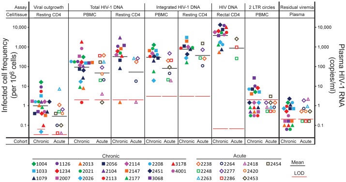
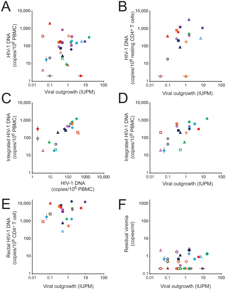
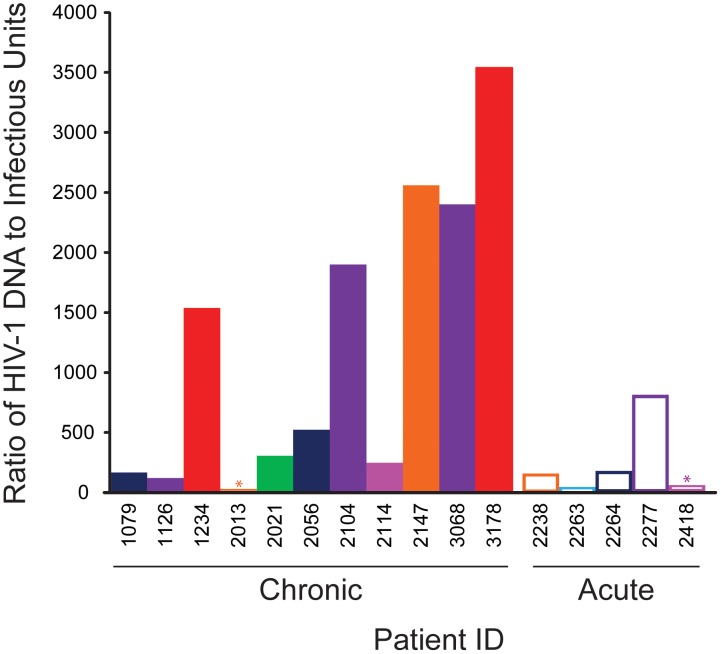
HIV-1 reservoirs preclude virus eradication in patients receiving highly active antiretroviral therapy (HAART). The best characterized reservoir is a small, difficult-to-quantify pool of resting memory CD4(+) T cells carrying latent but replication-competent viral genomes. Because strategies targeting this latent reservoir are now being tested in clinical trials, well-validated high-throughput assays that quantify this reservoir are urgently needed. Here we compare eleven different approaches for quantitating persistent HIV-1 in 30 patients on HAART, using the original viral outgrowth assay for resting CD4(+) T cells carrying inducible, replication-competent viral genomes as a standard for comparison. PCR-based assays for cells containing HIV-1 DNA gave infected cell frequencies at least 2 logs higher than the viral outgrowth assay, even in subjects who started HAART during acute/early infection. This difference may reflect defective viral genomes. The ratio of infected cell frequencies determined by viral outgrowth and PCR-based assays varied dramatically between patients. Although strong correlations with the viral outgrowth assay could not be formally excluded for most assays, correlations achieved statistical significance only for integrated HIV-1 DNA in peripheral blood mononuclear cells and HIV-1 RNA/DNA ratio in rectal CD4(+) T cells. Residual viremia was below the limit of detection in many subjects and did not correlate with the viral outgrowth assays. The dramatic differences in infected cell frequencies and the lack of a precise correlation between culture and PCR-based assays raise the possibility that the successful clearance of latently infected cells may be masked by a larger and variable pool of cells with defective proviruses. These defective proviruses are detected by PCR but may not be affected by reactivation strategies and may not require eradication to accomplish an effective cure. A molecular understanding of the discrepancy between infected cell frequencies measured by viral outgrowth versus PCR assays is an urgent priority in HIV-1 cure research.
Conflict of interest statement
The authors have declared that no competing interests exist.
Figures
References
-
- Gulick RM, Mellors JW, Havlir D, Eron JJ, Gonzalez C, et al. (1997) Treatment with indinavir, zidovudine, and lamivudine in adults with human immunodeficiency virus infection and prior antiretroviral therapy. N Engl J Med 337: 734–739. - PubMed
-
- Hammer SM, Squires KE, Hughes MD, Grimes JM, Demeter LM, et al. (1997) A controlled trial of two nucleoside analogues plus indinavir in persons with human immunodeficiency virus infection and CD4 cell counts of 200 per cubic millimeter or less. AIDS clinical trials group 320 study team. N Engl J Med 337: 725–733. - PubMed
-
- Perelson AS, Essunger P, Cao Y, Vesanen M, Hurley A, et al. (1997) Decay characteristics of HIV-1-infected compartments during combination therapy. Nature 387: 188–191. - PubMed
-
- Chun TW, Finzi D, Margolick J, Chadwick K, Schwartz D, et al. (1995) In vivo fate of HIV-1-infected T cells: Quantitative analysis of the transition to stable latency. Nat Med 1: 1284–1290. - PubMed
-
- Chun TW, Carruth L, Finzi D, Shen X, DiGiuseppe JA, et al. (1997) Quantification of latent tissue reservoirs and total body viral load in HIV-1 infection. Nature 387: 183–188. - PubMed
Publication types
MeSH terms
Substances
Grants and funding
- P30 MH062246/MH/NIMH NIH HHS/United States
- U19 AI096109/AI/NIAID NIH HHS/United States
- AI69432-S/AI/NIAID NIH HHS/United States
- UL 1 RR024131/RR/NCRR NIH HHS/United States
- AI306214/AI/NIAID NIH HHS/United States
- R37 AI051178/AI/NIAID NIH HHS/United States
- R01AIO87145/PHS HHS/United States
- 1U19AI096109/AI/NIAID NIH HHS/United States
- P30 AIO27763/PHS HHS/United States
- U19 AI096113/AI/NIAID NIH HHS/United States
- P30 MH62246/MH/NIMH NIH HHS/United States
- R24 AI106039/AI/NIAID NIH HHS/United States
- P01 AI080193/AI/NIAID NIH HHS/United States
- K24AIO69994/PHS HHS/United States
- T32 AI007632/AI/NIAID NIH HHS/United States
- IK2 CX000520/CX/CSRD VA/United States
- P30 DK026743/DK/NIDDK NIH HHS/United States
- K23 CA157929/CA/NCI NIH HHS/United States
- UM1 AI069432/AI/NIAID NIH HHS/United States
- AI080193/AI/NIAID NIH HHS/United States
- HHMI/Howard Hughes Medical Institute/United States
- K24 AI069994/AI/NIAID NIH HHS/United States
- U01 AI069432/AI/NIAID NIH HHS/United States
- AI74621/AI/NIAID NIH HHS/United States
- P30 AI027763/AI/NIAID NIH HHS/United States
- 43222/PHS HHS/United States
- P01 AI074621/AI/NIAID NIH HHS/United States
- AI096113/AI/NIAID NIH HHS/United States
LinkOut - more resources
Full Text Sources
Other Literature Sources
Medical
Research Materials
