Lumbosacral plexus injury following vaginal delivery with epidural analgesia -A case report-
- PMID: 23459069
- PMCID: PMC3581790
- DOI: 10.4097/kjae.2013.64.2.175
Lumbosacral plexus injury following vaginal delivery with epidural analgesia -A case report-
Abstract
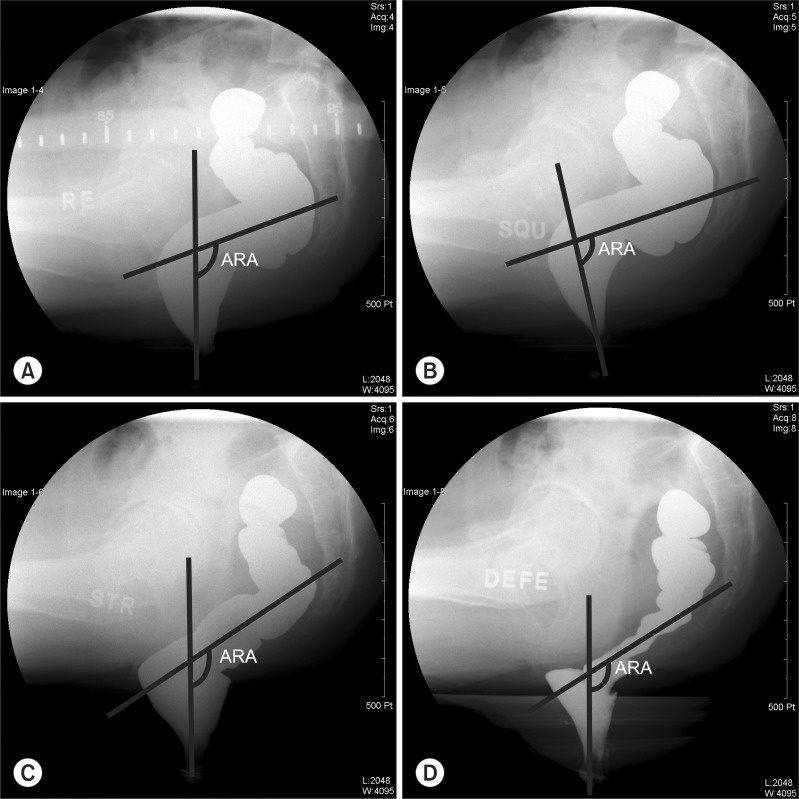
A 26 year old, healthy, 41 week primiparous woman received a patient-controlled epidural analgesia (PCEA) and experienced paraplegia 11 hours later after a vaginal delivery. This was thought to be the result of complications from PCEA but there was no specific abnormality on magnetic resonance imaging (MRI) of the lumbosacral spine. On an electromyography (EMG) study performed 15 days following delivery, signs of tibial neuropathy were present and peripheral nerve injury during vaginal delivery was suspected. Motor weakness and hypoesthesia of both lower extremities improved rapidly, but a decrease in the desire to urinate or defecate, followed by urinary incontinence and constipation persisted, We suspected the sacral plexus had been severely damaged during vaginal delivery. Seven months later, the patient's conditions improved but had not fully recovered.
Keywords: Analgesia; Epidural; Lumbosacral plexus; Obstetric delivery.
Figures


Similar articles
-
Postpartum lumbosacral radiculopathy: a neuraxial anaesthesia complication or an intrinsic obstetric palsy?BMJ Case Rep. 2021 Apr 21;14(4):e241669. doi: 10.1136/bcr-2021-241669. BMJ Case Rep. 2021. PMID: 33883118 Free PMC article.
-
Transient paraplegia due to accidental intrathecal bupivacaine infiltration following pre-emptive analgesia in a patient with missed sacral dural ectasia.Spine (Phila Pa 1976). 2010 Nov 15;35(24):E1444-6. doi: 10.1097/BRS.0b013e3181e91e2b. Spine (Phila Pa 1976). 2010. PMID: 21030892 Review.
-
Effects of Patient-Controlled Epidural Analgesia on Uterine Electromyography During Spontaneous Onset of Labor in Term Nulliparous Women.Reprod Sci. 2015 Nov;22(11):1350-7. doi: 10.1177/1933719115578926. Epub 2015 Mar 29. Reprod Sci. 2015. PMID: 25824008
-
Sciatic neuropathy after normal vaginal delivery: A case report.J Clin Neurosci. 2020 Feb;72:480-482. doi: 10.1016/j.jocn.2019.11.031. Epub 2019 Dec 9. J Clin Neurosci. 2020. PMID: 31822439
-
Spontaneous Spinal Epidural Hematoma After Normal Spontaneous Delivery with Epidural Analgesia: Case Report and Literature Review.World Neurosurg. 2020 May;137:214-217. doi: 10.1016/j.wneu.2020.01.240. Epub 2020 Feb 10. World Neurosurg. 2020. PMID: 32058108 Review.
Cited by
-
Postpartum lumbosacral radiculopathy: a neuraxial anaesthesia complication or an intrinsic obstetric palsy?BMJ Case Rep. 2021 Apr 21;14(4):e241669. doi: 10.1136/bcr-2021-241669. BMJ Case Rep. 2021. PMID: 33883118 Free PMC article.
-
Intrinsic Obstetric Palsy: Case Report and Literature Review.J Clin Diagn Res. 2016 Apr;10(4):QD06-7. doi: 10.7860/JCDR/2016/18797.7693. Epub 2016 Apr 1. J Clin Diagn Res. 2016. PMID: 27190901 Free PMC article.
-
Good prognosis of postpartum lower limb sensorimotor deficit: a combined clinical, electrophysiological, and radiological follow-up.J Neurol. 2017 Mar;264(3):529-540. doi: 10.1007/s00415-016-8388-5. Epub 2017 Jan 6. J Neurol. 2017. PMID: 28062970
References
-
- Wong CA, Scavone BM, Dugan S, Smith JC, Prather H, Ganchiff JN, et al. Incidence of postpartum lumbosacral spine and lower extremity nerve injuries. Obstet Gynecol. 2003;101:279–288. - PubMed
-
- Loo CC, Dahlgren G, Irestedt L. Neurological complications in obstetric regional anesthesia. Int J Obstet Anesth. 2000;9:99–124. - PubMed
-
- Moen V, Irestedt L. Neurological complications following central neuraxial blockades in obstetrics. Curr Opin Anaesthesiol. 2008;21:275–280. - PubMed
-
- Holdcroft A, Gibberd FB, Hargrove RL, Hawkins DF, Dellaportas CI. Neurological complications associated with pregnancy. Br J Anaesth. 1995;75:522–526. - PubMed
-
- Horlocker TT, Wedel DJ. Anticoagulation and neuraxial block: historical perspective, anesthetic implications, and risk management. Reg Anesth Pain Med. 1998;23(6 Suppl 2):129–134. - PubMed
LinkOut - more resources
Full Text Sources
Other Literature Sources