

Long-term effects of alendronate on fracture healing and bone remodeling of femoral shaft in ovariectomized rats
- PMID: 23459092
- PMCID: PMC4002493
- DOI: 10.1038/aps.2012.170
Long-term effects of alendronate on fracture healing and bone remodeling of femoral shaft in ovariectomized rats
Abstract
Aim: To investigate the long-term effects of alendronate (Aln), a widely used oral bisphosphonate, on fracture healing and bone remodeling in ovariectomized rats.
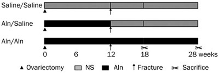
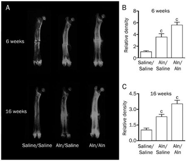
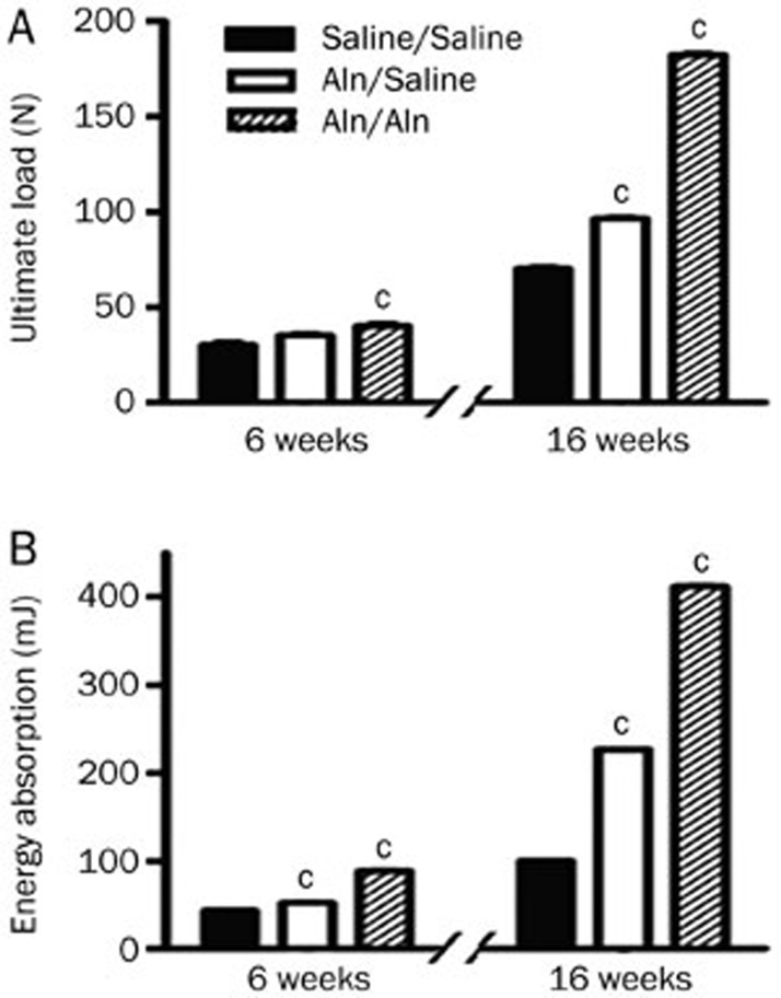
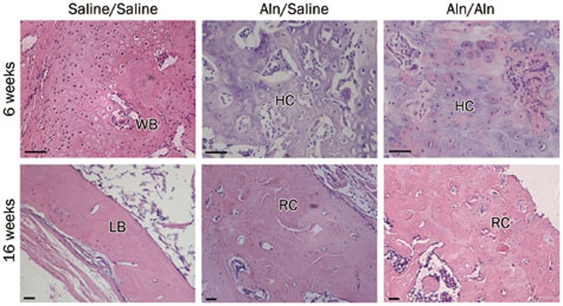
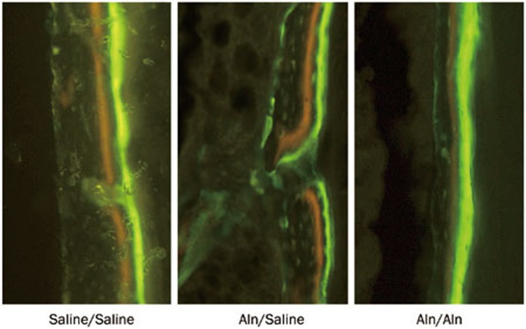
Methods: Adult female SD rats underwent ovariectomy, and then bilateral femoral osteotomy at 12 weeks post-ovariectomy. From d 2 post-ovariectomy, the animals were divided into 3 groups, and treated with Aln (3 mg·kg(-1)·d(-1), po) for 28 weeks (Aln/Aln), Aln for 12 weeks and saline for 16 weeks (Aln/Saline) or saline for 28 weeks (Saline/Saline). At 6 and 16 weeks post-fracture, the fracture calluses were examined with X-ray radiography, and biomechanical testing and histological analysis were performed. The calluses were labeled with tetracycline and calcein to evaluate the mineral apposition rate (MAR).
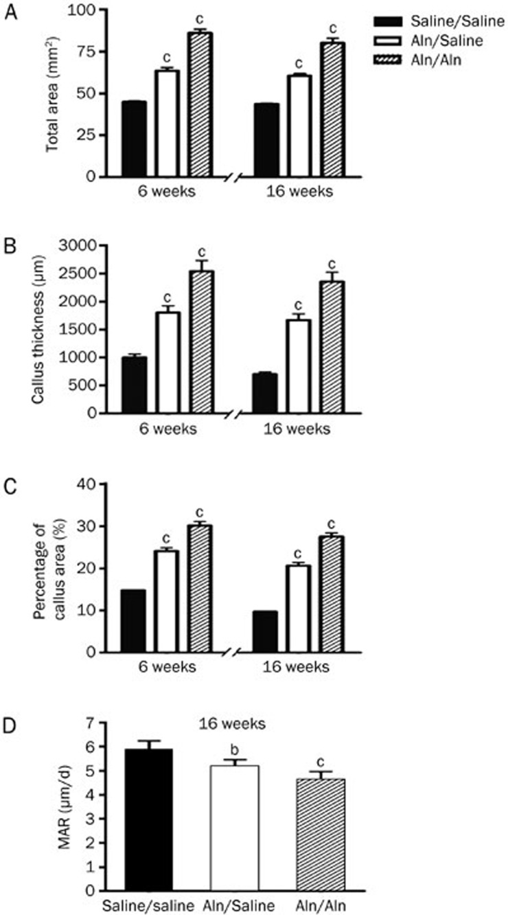
Results: The fracture line was less distinct in the 2 Aln-treated groups at 6 weeks post-fracture, and disappeared in all the 3 groups at 16 weeks post-fracture. The size of the callus and radiographic density of the femora in the Aln/Aln group were the highest among the 3 groups at 6 and 16 weeks post-fracture. Similar results were observed in the ultimate load at failure and energy absorption. However, the treatment with Aln delayed endochondral ossification of the callus, and significantly increased the total sagittal-sectional area, percentage callus area and callus thickness, and decreased the MAR at 6 and 16 weeks post-fracture.
Conclusion: In the ovariectomized rat model, Aln is beneficial for the mechanical properties of the callus, but delays callus remodeling by suppressing the remodeling of woven bone into lamellar bone.
Figures
References
-
- Giannoudis P, Tzioupis C, Almalki T, Buckley R.Fracture healing in osteoporotic fractures: is it really different? A basic science perspective Injury 200738Suppl 1: S90–9. - PubMed
-
- Geusens P. Bisphosphonates for postmenopausal osteoporosis: determining duration of treatment. Curr Osteoporos Rep. 2009;7:12–7. - PubMed
-
- Müller D, Pulm J, Gandjour A. Cost-effectiveness of different strategies for selecting and treating individuals at increased risk of osteoporosis or osteopenia: a systematic review. Value Health. 2012;15:284–98. - PubMed
-
- Fu L, Tang T, Miao Y, Zhang S, Qu Z, Dai K. Stimulation of osteogenic differentiation and inhibition of adipogenic differentiation in bone marrow stromal cells by alendronate via ERK and JNK activation. Bone. 2008;43:40–7. - PubMed
-
- Schneider JP. Bisphosphonates and low-impact femoral fractures: current evidence on alendronate-fracture risk. Geriatrics. 2009;64:18–23. - PubMed
Publication types
MeSH terms
Substances
LinkOut - more resources
Full Text Sources
Other Literature Sources
Medical
