

Biomechanical factors in the biology of aortic wall and aortic valve diseases
- PMID: 23459103
- PMCID: PMC3695745
- DOI: 10.1093/cvr/cvt040
Biomechanical factors in the biology of aortic wall and aortic valve diseases
Abstract
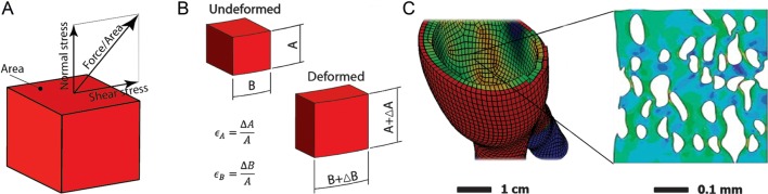
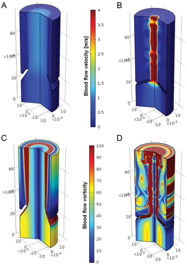
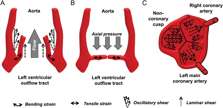
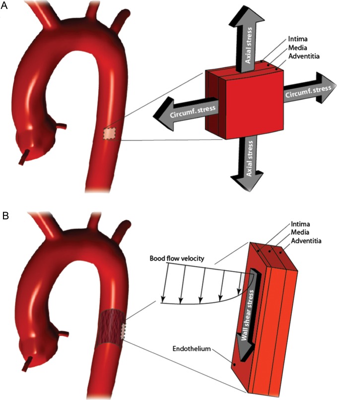
The biomechanical factors that result from the haemodynamic load on the cardiovascular system are a common denominator of several vascular pathologies. Thickening and calcification of the aortic valve will lead to reduced opening and the development of left ventricular outflow obstruction, referred to as aortic valve stenosis. The most common pathology of the aorta is the formation of an aneurysm, morphologically defined as a progressive dilatation of a vessel segment by more than 50% of its normal diameter. The aortic valve is exposed to both haemodynamic forces and structural leaflet deformation as it opens and closes with each heartbeat to assure unidirectional flow from the left ventricle to the aorta. The arterial pressure is translated into tension-dominated mechanical wall stress in the aorta. In addition, stress and strain are related through the aortic stiffness. Furthermore, blood flow over the valvular and vascular endothelial layer induces wall shear stress. Several pathophysiological processes of aortic valve stenosis and aortic aneurysms, such as macromolecule transport, gene expression alterations, cell death pathways, calcification, inflammation, and neoangiogenesis directly depend on biomechanical factors.
Keywords: Abdominal aortic aneurysm; Aortic stenosis; Inflammation; Thoracic aortic aneurysm.
Figures
References
-
- Dobzhansky T. Nothing in biology makes sense except in the light of evolution. Am Biol Teach. 1973;5:125–129. doi:10.2307/4444260. - DOI
-
- Michel JB, Martin-Ventura JL, Egido J, Sakalihasan N, Treska V, Lindholt J, et al. Novel aspects of the pathogenesis of aneurysms of the abdominal aorta in humans. Cardiovasc Res. 2011;90:18–27. doi:10.1093/cvr/cvq337. - DOI - PMC - PubMed
-
- Yang N, Vafai K. Low-density lipoprotein (LDL) transport in an artery – A simplified analytical solution. Int J Heat Mass Transfer. 2008;51:497–505. doi:10.1016/j.ijheatmasstransfer.2007.05.023. - DOI
-
- Caro CG. Discovery of the role of wall shear in atherosclerosis. Arterioscler Thromb Vasc Biol. 2009;29:158–161. doi:10.1161/ATVBAHA.108.166736. - DOI - PubMed
-
- Rajamannan NM, Evans FJ, Aikawa E, Grande-Allen KJ, Demer LL, Heistad DD, et al. Calcific aortic valve disease: not simply a degenerative process. Circulation. 2011;124:1783–1791. doi:10.1161/CIRCULATIONAHA.110.006767. - DOI - PMC - PubMed
Publication types
MeSH terms
LinkOut - more resources
Full Text Sources
Other Literature Sources
Medical
