. 2013;7(2):e2048.
doi: 10.1371/journal.pntd.0002048. Epub 2013 Feb 14.

Development and validation of a novel diagnostic test for human brucellosis using a glyco-engineered antigen coupled to magnetic beads

Affiliations

Development and validation of a novel diagnostic test for human brucellosis using a glyco-engineered antigen coupled to magnetic beads

Andrés E Ciocchini et al. PLoS Negl Trop Dis. 2013.

Abstract

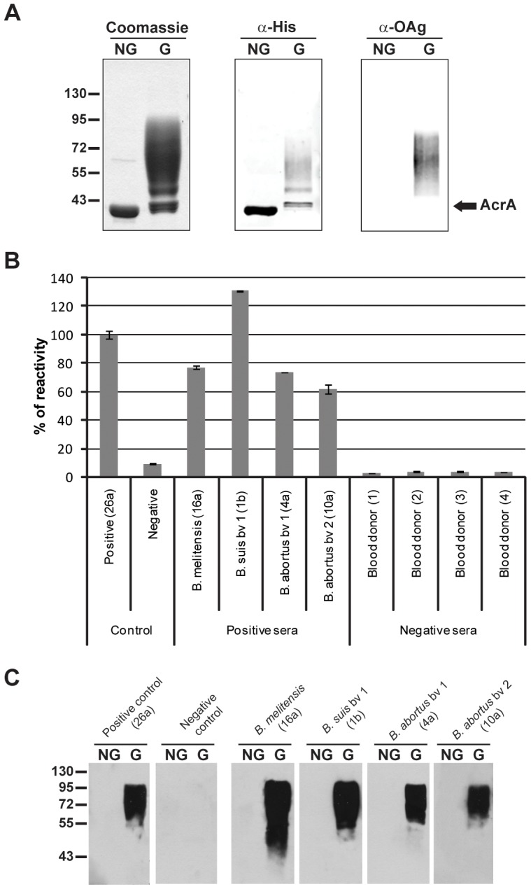
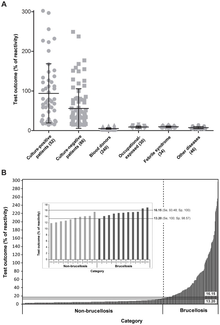
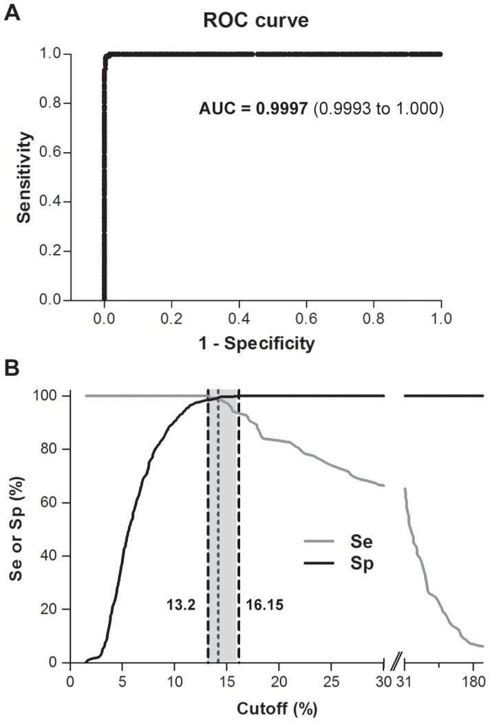
Brucellosis is a highly contagious zoonosis and still a major human health problem in endemic areas of the world. Although several diagnostic tools are available, most of them are difficult to implement especially in developing countries where complex health facilities are limited. Taking advantage of the identical structure and composition of the Brucella spp. and Yersinia enterocolitica O:9 O-polysaccharide, we explored the application of a recombinant Y. enterocolitica O:9-polysaccharide-protein conjugate (OAg-AcrA) as a novel antigen for diagnosis of human brucellosis. We have developed and validated an indirect immunoassay using OAg-AcrA coupled to magnetic beads. OAg-AcrA was produced and purified with high yields in Y. enterocolitica O:9 cells co-expressing the oligosaccharyltransferase PglB and the protein acceptor AcrA of Campylobacter jejuni without the need for culturing Brucella. Expression of PglB and AcrA in Y. enterocolitica resulted in the transfer of the host O-polysaccharide from its lipid carrier to AcrA. To validate the assay and determine the cutoff values, a receiver-operating characteristic analysis was performed using a panel of characterized serum samples obtained from healthy individuals and patients of different clinical groups. Our results indicate that, using this assay, it is possible to detect infection caused by the three main human brucellosis agents (B. abortus, B. melitensis and B. suis) and select different cutoff points to adjust sensitivity and specificity levels as needed. A cutoff value of 13.20% gave a sensitivity of 100% and a specificity of 98.57%, and a cutoff value of 16.15% resulted in a test sensitivity and specificity of 93.48% and 100%, respectively. The high diagnostic accuracy, low cost, reduced assay time and simplicity of this new glycoconjugate-magnetic beads assay makes it an attractive diagnostic tool for using not only in clinics and brucellosis reference laboratories but also in locations with limited laboratory infrastructure and/or minimally trained community health workers.


Conflict of interest statement

A provisional patent has been filed regarding the diagnostic application of recombinant glycoconjugates.

Figures

Figure 1
Figure 1. OAg-AcrA glycoconjugate as an antigen for the diagnosis of human brucellosis.
A) 10% SDS-PAGE analysis of purified non-glycosylated (NG) and glycosylated AcrA (G) by Coomassie brilliant blue and immunoblot using anti-histidine tag and anti-Brucella O-antigen monoclonal antibodies. B) Glycoconjugate magnetic beads-based immunoassay for detection of antibodies against the O-polysaccharide fraction of lipopolysaccharide (LPS). Magnetic beads coated with OAg-AcrA were incubated with the indicated human serum samples. Bound antibodies were detected using Cy5-conjugated goat anti human IgM/G antibodies.Positive and negative controls; human sera included as positive and negative controls in each assay run. The figures in parenthesis correspond to the identification of the samples according to Tables S1 and S2. Results are expressed as percentage of reactivity of the positive control serum. The bar graph data represents the means and standard deviation for two separate determinations. C) Western blot analysis of the same human serum samples.
Figure 2
Figure 2. Glycoconjugate-magnetic beads assay analysis of serum samples obtained from healthy individuals and patients of different clinical groups.
A) Dotplot of the glycoconjugate-magnetic beads assay results. Serum samples of blood culture-positive patients (52 sera), culture-negative/serologically-positive patients with clinical diagnosis of brucellosis (86 sera), blood donors (240 sera), healthy individuals occupational-exposed to the pathogen (30 sera), patients with febrile syndrome (34 sera) and patients with other diseases (46 sera) were tested as indicated in Materials and Methods. The mean and standard deviation for each group are indicated. B) Analysis of glycoconjugate-magnetic beads assay results classifying the samples into two categories. Non-brucellosis category includes samples obtained from blood donors, healthy individuals occupational-exposed to the pathogen, patients with febrile syndrome and patients with other diseases. Brucellosis category includes samples of blood culture-positive and culture-negative/serologically-positive patients with clinical diagnosis of brucellosis. For each category reactivity values are arrange in increasing order. The horizontal lines indicate the cutoff values calculated by ROC analysis for which maximal sensitivity (Se) or specificity (Sp) is achieved. The inset shows a zoom of the graph part displaying the limit between categories.
Figure 3
Figure 3. Receiver-operating characteristic (ROC) analysis of glycoconjugate-magnetic beads assay results.
A) ROC plot. The analysis was carried out considering as positive controls sera of patients with culture-confirmed brucellosis and culture-negative/serologically-positive patients with clinical diagnosis of brucellosis (138 sera), and as negative controls serum samples from blood-donors, healthy individuals occupational-exposed to the pathogen, patients with febrile syndrome and patients with other diseases (350 sera). AUC, area under the ROC curve. Values in parentheses indicate the 95% confidence interval. B) Plot of the diagnostic sensitivity (Se) and specificity (Sp) of the assay as a function of the cutoff value. The dot vertical line indicates the cutoff value that concurrently optimizes Se and Sp (cutoff = 14.18%, Se = 99.28% and Sp = 99.43%). The dash vertical lines indicate the cutoff values for which maximal Se or Sp is achieved (cutoff = 13.20%, Se = 100% and Sp = 98.57%; cutoff = 16.15%, Se = 93.48% and Sp = 100%).

References

    1. Pappas G, Papadimitriou P, Akritidis N, Christou L, Tsianos EV (2006) The new global map of human brucellosis. Lancet Infect Dis 6: 91–99. - PubMed
    1. Franco MP, Mulder M, Gilman RH, Smits HL (2007) Human brucellosis. Lancet Infect Dis 7: 775–786. - PubMed
    1. Young EJ (1995) An overview of human brucellosis. Clin Infect Dis 21: 283–289 quiz 290. - PubMed
    1. Al Dahouk S, Nockler K (2011) Implications of laboratory diagnosis on brucellosis therapy. Expert Rev Anti Infect Ther 9: 833–845. - PubMed
    1. Araj GF (2010) Update on laboratory diagnosis of human brucellosis. Int J Antimicrob Agents 36 Suppl 1: S12–17. - PubMed

Publication types