

Role of ultrasound and color Doppler imaging in the detection of carotid paragangliomas
- PMID: 23459221
- PMCID: PMC3558042
- DOI: 10.1016/j.jus.2012.05.001
Role of ultrasound and color Doppler imaging in the detection of carotid paragangliomas
Abstract
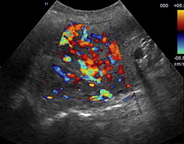
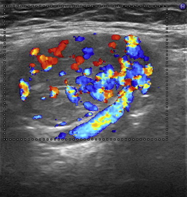
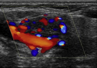
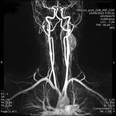
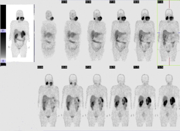
Introduction: Carotid body paragangliomas (PGLs) are highly vascularized lesions that arise from the paraganglia located at the carotid bifurcation.
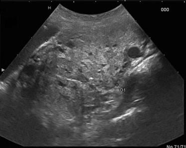
Purpose: To evaluate the usefulness of gray-scale ultrasound (US) and color Doppler ultrasound (CDUS) in the detection and follow-up of carotid PGLs of the neck.
Materials and methods: The authors retrospectively reviewed US and CDUS examinations of the neck performed in 40 patients with PGL syndrome type 1 and single or bilateral neck PGLs confirmed by CT or MRI; the patients had a total of 60 PGLs of the neck. US and CDUS outcome was compared to the outcome of second-line imaging techniques such as magnetic resonance imaging (MRI) or computed tomography (CT). The following findings were considered: presence/absence of focal lesions at US imaging and difference in maximum diameter of the lesion measured at US and MRI/CT. Results were compared using the Student's t-test.
Results: Of the 60 PGLs of the neck only 5 (8.3%) were not visualized at US or CDUS examination. The difference in maximum diameter of these lesions measured at CT/MRI and US/CDUS ranged between -5 mm and +16 mm (mean difference 2.2 ± 6.0). This difference was statistically significant (p = 0.008).
Conclusions: US and CDUS are useful methods for identifying carotid PGLs also measuring less than 10 mm in diameter. However, diagnostic accuracy of US and CDUS is reduced in the measurement of the exact dimensions of the lesions.
Introduzione: I paragangliomi (PGLs) carotidei sono lesioni altamente vascolarizzate che originano dai paragangli localizzati a livello della biforcazione carotidea.
Scopo: Valutare l’utilità dell’ecografia (US) e dell’Eco color Doppler (USD) del collo nella diagnosi e nel follow-up dei PGLs carotidei.
Materiali e metodi: Abbiamo visionato retrospettivamente tutte le US e gli USD del collo, eseguiti nell’Ospedale Santa Chiara di Trento tra il 2007 e il 2011, di soggetti affetti da sindrome paraganglioma di tipo 1 con sicuri paragangliomi del collo singoli o bilaterali. Abbiamo quindi confrontato i risultati con quelli di metodiche di imaging di secondo livello, Risonanza Magnetica (RM) o Tomografia Computerizzata (TC). Sono stati calcolati i casi discordanti in termini di presenza/assenza della lesione focale alle immagini ecografiche e in termini di differenze del diametro maggiore rilevate tra le due metodiche.
Risultati: Sono stati revisionati i dati di imaging (US e/o USD, RM o TC) eseguiti tra il 2007 e il 2011 di 40 pazienti aventi 60 sicuri paragangliomi del collo. Di questi solo 5/60 (8,3%) non sono stati visualizzati mediante US e/o USD. La differenza nella misura del diametro maggiore di tali lesioni rilevate dalle due tecniche di imaging, è risultata compresa tra -5 e +16 mm (media di 2,2 ± 6,0). Tale differenza è risultata statisticamente significativa (p = 0,008) (test-Student).
Conclusioni: L’US e l’USD del collo risultano metodiche di imaging utili nell’identificazione dei PGL carotidei anche in caso di dimensioni inferiori al centimetro. La loro accuratezza diagnostica si riduce se consideriamo le misure esatte delle lesioni.
Keywords: Carotid body paraganglioma; Carotid-body tumor; Color Doppler ultrasound; Ultrasound.
Figures
References
-
- Najibi S., Terramani T.T., Brinkman W., Thourani V.H., Smith R.B., 3rdm, Lumsden A.B. Carotid body tumors. J Am Coll Surg. 2002;194:538–539. - PubMed
-
- Schiavi F., Demattè S., Cecchini M.E., Taschin E., Bobisse S., Del Piano A. The endemic paraganglioma syndrome type 1: origin, spread, and clinical expression. J Clin Endocrinol Metab. 2012;97(4):E637–E641. - PubMed
-
- Martin T.P. What we call them: the nomenclature of head and neck paragangliomas. Clin Otolaryngol. 2006;31:185–186. - PubMed
-
- Grotemeyer D., Loghmanieh S.M., Pourhassan S., Sagban T.A., Iskandar F., Reinecke P. Dignity of carotid body tumors. Review of the literature and clinical experiences. Chirurg. 2009;80:854–863. - PubMed
-
- Lack E.E., Cubilla A.L., Woodruff J.M., Farr H.W. Paragangliomas of the head and neck region. A clinical study of 69 patients. Cancer. 1977;39:397–409. - PubMed
LinkOut - more resources
Full Text Sources
Miscellaneous