Review
. 2013 Jun-Jul;1829(6-7):514-22.
doi: 10.1016/j.bbagrm.2013.02.011. Epub 2013 Feb 28.

The social fabric of the RNA degradosome

Affiliations
Review

The social fabric of the RNA degradosome

Katarzyna J Bandyra et al. Biochim Biophys Acta. 2013 Jun-Jul.

Abstract

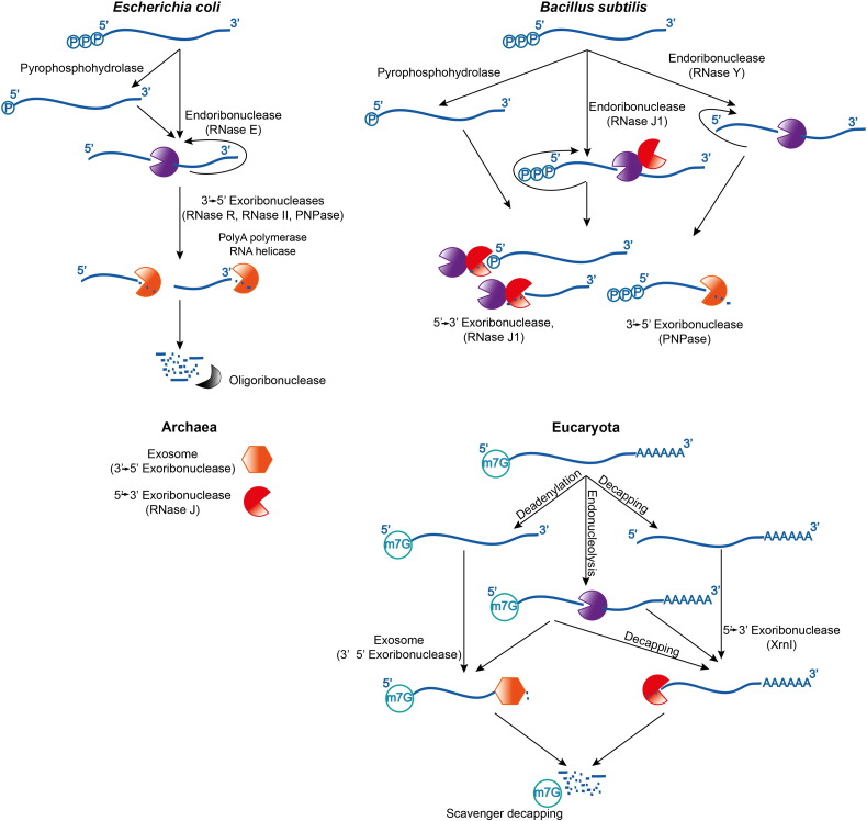
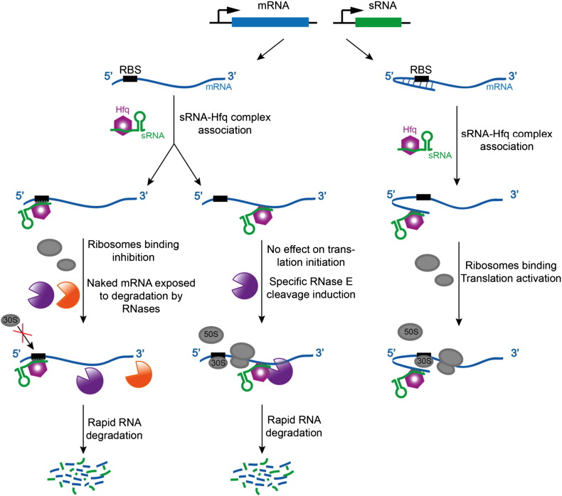
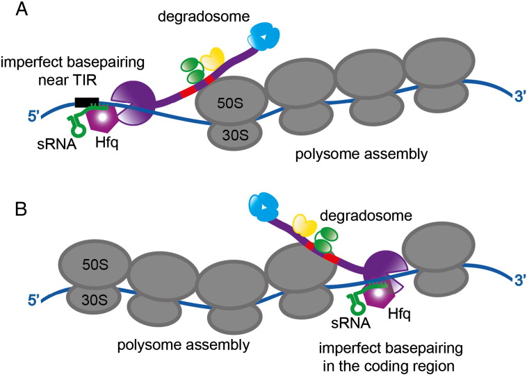
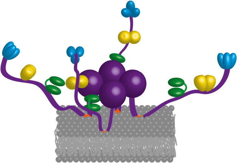
Bacterial transcripts each have a characteristic half-life, suggesting that the processes of RNA degradation work in an active and selective manner. Moreover, the processes are well controlled, thereby ensuring that degradation is orderly and coordinated. Throughout much of the bacterial kingdom, RNA degradation processes originate through the actions of assemblies of key RNA enzymes, known as RNA degradosomes. Neither conserved in composition, nor unified by common evolutionary ancestry, RNA degradosomes nonetheless can be found in divergent bacterial lineages, implicating a common requirement for the co-localisation of RNA metabolic activities. We describe how the cooperation of components in the representative degradosome of Escherichia coli may enable controlled access to transcripts, so that they have defined and programmable lifetimes. We also discuss how this cooperation contributes to precursor processing and to the riboregulation of intricate post-transcriptional networks in the control of gene expression. The E. coli degradosome interacts with the cytoplasmic membrane, and we discuss how this interaction may spatially organise the assembly and contribute to subunit cooperation and substrate capture. This article is part of a Special Issue entitled: RNA Decay mechanisms.


Figures

Fig. 1
Fig. 1
Overview of RNA decay pathways in the different life domains. Within the two main bacterial lineages, represented by the gram-negative Escherichia coli and the gram-positive Bacillus subtilis (top left and top right panels, respectively), the enzymes differ although the pathways share similarity. In both gram-negative and gram-positive lineages, the endoribonucleases can cleave the substrates repetitively, and the products are attacked by exoribonucleases. The degradation pathways in eukaryotes are shown for comparison.
Fig. 2
Fig. 2
Components of the canonical degradosome. The asterisk marks the predicted size of the catalytic domain of RNase E; the amino acids 511–529 were not visible in the crystal structure .
Fig. 3
Fig. 3
The quaternary structure of RNase E and its modes of structural change: A) Structural domains of RNase E (color coded) with a 13mer RNA oligo bound (showed only in one protomer for clarity) and the quaternary structural organisation. The tetramer is a dimer-of-dimers, and the principal dimer pairs are at the same horizontal level and are linked by a zinc ion (small grey sphere). B) The 5′ phosphate binding pocket of RNase E with the RNA bound; the main amino acids contacting the phosphate group are labelled. C) The RNA substrate tracking along a shallow groove at the active site, with the contacts between RNA and RNase E marked in black. D) Domain movement in the RNase E catalytic domain upon substrate binding, and quaternary structure flexibility. The left panel shows the overall structure of RNase E tetramer alone (top) and the RNA bound (bottom). The movements at the dimer-of-dimer interface are shown in the middle panel. Domain closure with the S1/5′-sensor domain clamping down on the substrate is demonstrated on the right panel.
Fig. 4
Fig. 4
A cartoon schematic of two pathways for RNA cleavage by RNase E and the degradosome: 5′ end sensing and the 5′ bypass pathway. A). Activation of RNase E by 5′ end sensing to trigger domain closure. B). Proposed model for how the CTD might affect 5′ end bypass by capturing and presenting potential substrates to the catalytic domain. The red bars represent the two RNA binding domains in the C-terminal half of RNase E.
Fig. 5
Fig. 5
RNase E, the degradosome and sRNA mediated post-transcriptional regulation. Cartoon schematic of the sRNA mediated pathways for degradation (left and central panels) and translational activation (right panel). The purple body represents RNase E and the orange body is an exoribonuclease.
Fig. 6
Fig. 6
A speculative model for interaction of the degradosome and polysome in sRNA mediated gene silencing . Panel A suggests a mechanism for sRNA mediated activation of RNase E on the 5′ end of a spooling transcript. B) A more radical “emergency stop” process to terminate translation prematurely. Here, the cleavage is mediated in the coding region of an actively translated mRNA. This hypothetical mode would generate a stop-less transcript and require collaboration with the tmRNA pathway to rescue the terminated assembly. RNase E is purple, and the two RNA binding sites in the C-terminal half of the enzyme are shown in red. Helicase is the bi-lobed green body, enolase is the yellow dimer and PNPase is the blue trimer.
Fig. 7
Fig. 7
The membrane localisation of Escherichia coli RNase E and the RNA degradosome. Schematic of proposed association of an RNase E tetramer (purple) on the inner leaflet of the cytoplasmic membrane through four amphipathic alpha helices (orange). With this model, the hydrophobic side chains of the amphipathic helices are partially immersed in the hydrocarbon interior of the lipid bilayer (gray lines). The grey spheres are the lipid polar head groups. Membrane association may help to bring the other degradosome components closer in space. Each purple sphere is an RNase E catalytic domain, RhlB helicase is the bi-lobed green body, dimeric enolase is shown in yellow and trimeric PNPase in blue.

References

    1. Dressaire C., Picard F., Redon E., Loubière P., Queinnec I., Girbal L., Cocaign-Bousquet M. Role of mRNA stability during bacterial adaptation. PloS One. 2013;8:e59059. - PMC - PubMed
    1. Miller C., Schwalb B., Maier K., Schulz D., Dumcke S., Zacher B., Mayer A., Sydow J., Marcinowski L., Dolken L., Martin D.E., Tresch A., Cramer P. Dynamic transcriptome analysis measures rates of mRNA synthesis and decay in yeast. Mol. Syst. Biol. 2011;7:458. - PMC - PubMed
    1. Richards J., Sundermeier T., Svetlanov A., Karzai A.W. Quality control of bacterial mRNA decoding and decay. Biochim. Biophys. Acta. 2008;1779:574–582. - PMC - PubMed
    1. Selinger D.W., Saxena R.M., Cheung K.J., Church G.M., Rosenow C. Global RNA half-life analysis in Escherichia coli reveals positional patterns of transcript degradation. Genome Res. 2003;13:216–223. - PMC - PubMed
    1. Carpousis A.J. The RNA degradosome of Escherichia coli: an mRNA-degrading machine assembled on RNase E. Annu. Rev. Microbiol. 2007;61:71–87. - PubMed

Publication types

MeSH terms

LinkOut - more resources