. 2013;7(2):e2071.
doi: 10.1371/journal.pntd.0002071. Epub 2013 Feb 14.

Placental histopathological changes associated with Plasmodium vivax infection during pregnancy

Affiliations

Placental histopathological changes associated with Plasmodium vivax infection during pregnancy

Rodrigo M Souza et al. PLoS Negl Trop Dis. 2013.

Erratum in

  • PLoS Negl Trop Dis. 2013 Apr;7(4). doi: 10.1371/annotation/28901e80-13ad-4cae-99e8-8d54625743b6. Epiphânio, Sabrina [corrected to Epiphanio, Sabrina]

Abstract

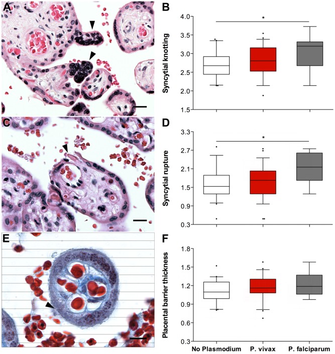
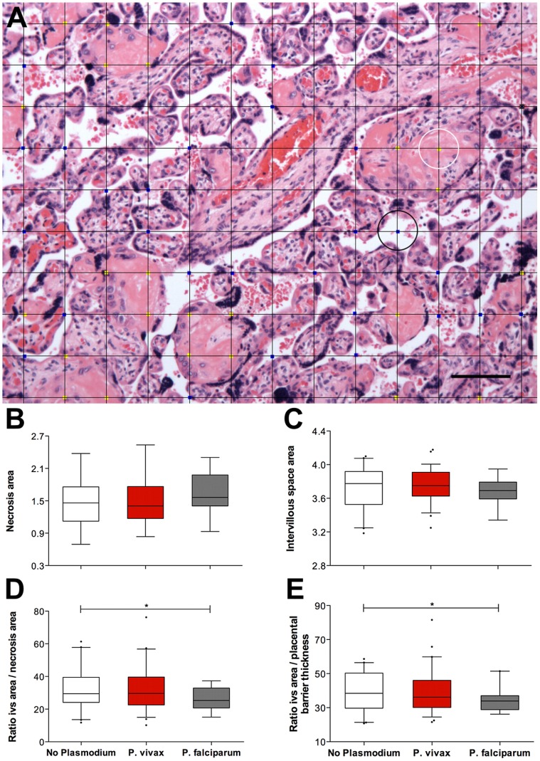
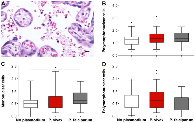
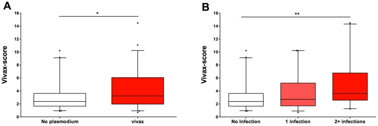
Histological evidence of Plasmodium in the placenta is indicative of placental malaria, a condition associated with severe outcomes for mother and child. Histological lesions found in placentas from Plasmodium-exposed women include syncytial knotting, syncytial rupture, thickening of the placental barrier, necrosis of villous tissue and intervillositis. These histological changes have been associated with P. falciparum infections, but little is known about the contribution of P. vivax to such changes. We conducted a cross-sectional study with pregnant women at delivery and assigned them to three groups according to their Plasmodium exposure during pregnancy: no Plasmodium exposure (n = 41), P. vivax exposure (n = 59) or P. falciparum exposure (n = 19). We evaluated their placentas for signs of Plasmodium and placental lesions using ten histological parameters: syncytial knotting, syncytial rupture, placental barrier thickness, villi necrosis, intervillous space area, intervillous leucocytes, intervillous mononucleates, intervillous polymorphonucleates, parasitized erythrocytes and hemozoin. Placentas from P. vivax-exposed women showed little evidence of Plasmodium or hemozoin but still exhibited more lesions than placentas from women not exposed to Plasmodium, especially when infections occurred twice or more during pregnancy. In the Brazilian state of Acre, where diagnosis and primary treatment are readily available and placental lesions occur in the absence of detected placental parasites, relying on the presence of Plasmodium in the placenta to evaluate Plasmodium-induced placental pathology is not feasible. Multivariate logistic analysis revealed that syncytial knotting (odds ratio [OR], 4.21, P = 0.045), placental barrier thickness (OR, 25.59, P = 0.021) and mononuclear cells (OR, 4.02, P = 0.046) were increased in placentas from P. vivax-exposed women when compared to women not exposed to Plasmodium during pregnancy. A vivax-score was developed using these three parameters (and not evidence of Plasmodium) that differentiates between placentas from P. vivax-exposed and unexposed women. This score illustrates the importance of adequate management of P. vivax malaria during pregnancy.


Conflict of interest statement

The authors have declared that no competing interests exist.

Figures

Figure 1
Figure 1. The syncytial parameters evaluated by Plasmodium species during infection.
Syncytial knotting (A and B) and syncytial rupture (C and D) were evaluated on H&E-stained slides at 100× magnification. Placental barrier thickness (E and F) was evaluated on Masson's trichrome-stained slides at 1000× magnification after overlaying horizontal lines with 5 µm of interspace (see Methods and Table 1). For all parameters, placentas from the “no plasmodium” group (n = 41; white boxes) had the lowest values, followed by placentas from the “P. vivax” (n = 59; red boxes) and “P. falciparum” (n = 19; grey boxes) groups. Graphs (B, D and F) represent the transformed data. * ANOVA test, P-value≤0,006. The boxes represent the mean and standard deviation values. The whiskers represent the 5th and 95th percentiles. Photographs were taken using a Zeiss Axio Imager M2 light microscope equipped with a Zeiss Axio Cam HRc. The grid overlays and counts were conducted using Image J. Arrow heads on A, C and D point to syncytial knots, syncytial rupture and an example of a thickness measurement, respectively.
Figure 2
Figure 2. The placental parameters evaluated by Plasmodium species during infection.
For all placentas, areas of necrosis (B) and intervillous space (C) were measured by overlaying a square grid (A) and counting the number of intersecting points that touched necrotic areas (yellow dots; the white circle indicates an example) or intervillous space areas (blue dots; the black circle indicates an example). The ratios of intervillous space area per necrosis (D) and intervillous space area per placental barrier thickness (E) were calculated. The placentas in the “no plasmodium” group (n = 41; white boxes) appear to have similar necrotic areas and more intervillous space than the placentas in the “P. vivax” group (n = 59; red boxes). The placentas in the “P. falciparum” group (n = 19; grey boxes) exhibited more necrotic areas and less intervillous space. Graphs (B, C, D and E) represent the transformed data. The boxes represent the mean and standard deviation values. The whiskers represent the 5th and 95th percentiles. The photograph was taken using a Zeiss Axio Imager M2 light microscope equipped with a Zeiss Axio Cam HRc. Grid overlays and counts were performed using Image J.
Figure 3
Figure 3. The immune-cell parameters evaluated by Plasmodium species during infection.
The percentage of immune cells present in the intervillous space of the placentas evaluated (A) was calculated after counting a total of 500 intervillous space cells. Total leucocytes percentage (B), mononuclear cells percentage (C) and polymorphonuclear cells percentage (D) were plotted against Plasmodium exposure during pregnancy, assessed by microscopy. The placentas from the “no plasmodium” group (n = 41; white boxes) appear to have less immune cells present in the intervillous space than the placentas from the “P. vivax” group (n = 59; red boxes) and the placentas from the “P. falciparum” group (n = 19; grey boxes). * ANOVA test, P-value = 0,039. Graphs (B, C, and D) represent the transformed data. The boxes represent the mean and standard deviation values. The whiskers represent the 5th and 95th percentiles. The photograph was taken using a Zeiss Axio Imager M2 light microscope equipped with a Zeiss Axio Cam HRc. Grid overlays and counts were performed using Image J.
Figure 4
Figure 4. The placental score differentiates the women who were exposed to P. vivax during pregnancy.
A score (termed the ‘vivax-score’) was developed and applied to all of the placental samples in this study (see main text for details). (A) The placental samples from the “no Plasmodium” group (n = 41, white box) revealed a significantly lower score than the placentas from the “P. vivax” group (n = 59, red box) (* Mann-Whitney, P = 0.027). (B) The vivax-score increased significantly (** Cuzick's trend test: z = 2.76, P = 0.006) with increased exposure to P. vivax during pregnancy. “No infection”, n = 41; “1 infection”, n = 39; “2+ infections”, n = 20).

References

    1. Dellicour S, Tatem AJ, Guerra Ca, Snow RW, Ter Kuile FO (2010) Quantifying the number of pregnancies at risk of malaria in 2007: a demographic study. PLoS Medicine 7: 1–10. - PMC - PubMed
    1. Steketee RW, Nahlen BL, Parise ME, Menendez C (2001) The burden of malaria in pregnancy in malaria-endemic areas. The American journal of tropical medicine and hygiene 64: 28–35. - PubMed
    1. Rogerson SJ, Hviid L, Duffy PE, Leke RF, Taylor DW (2007) Malaria in pregnancy: pathogenesis and immunity. Lancet Infect Dis 7: 105–117. - PubMed
    1. Menendez C (2006) Malaria during pregnancy. Current Molecular Medicine 6: 269–273. - PubMed
    1. Nosten F, McGready R, Simpson JA, Thwai KL, Balkan S, et al. (1999) Effects of Plasmodium vivax malaria in pregnancy. Lancet 354: 546–549. - PubMed

Publication types