

Automated Delineation of Lung Tumors from CT Images Using a Single Click Ensemble Segmentation Approach
- PMID: 23459617
- PMCID: PMC3580869
- DOI: 10.1016/j.patcog.2012.10.005
Automated Delineation of Lung Tumors from CT Images Using a Single Click Ensemble Segmentation Approach
Abstract
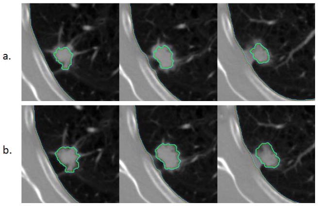
A single click ensemble segmentation (SCES) approach based on an existing "Click&Grow" algorithm is presented. The SCES approach requires only one operator selected seed point as compared with multiple operator inputs, which are typically needed. This facilitates processing large numbers of cases. Evaluation on a set of 129 CT lung tumor images using a similarity index (SI) was done. The average SI is above 93% using 20 different start seeds, showing stability. The average SI for 2 different readers was 79.53%. We then compared the SCES algorithm with the two readers, the level set algorithm and the skeleton graph cut algorithm obtaining an average SI of 78.29%, 77.72%, 63.77% and 63.76% respectively. We can conclude that the newly developed automatic lung lesion segmentation algorithm is stable, accurate and automated.
Keywords: CT; Delineation; Ensemble Segmentation; Image Features; Lesion; Lung Tumor; Region growing.
Figures


References
-
- Johnson DH, Blot WJ, Carbone DP. Cancer of the lung: Non-small cell lung cancer and small cell lung cancer. In: Abeloff MD, Armitage JO, Niederbuber JE, Kastan MB, McKenna WG, editors. Abeloff’s clinical oncology. Churchill Livingstone/Elsevier; Philadelphia: 2008.
-
- Rexilius J, Hahn HK, Schluter M, Bourquain H, Peitgen HO. Evaluation of accuracy in MS lesion volumetry using realistic lesion phantoms. Acad Radiol. 2005;12:17–24. - PubMed
-
- Tai P, Van Dyk J, Yu E, Battista J, Stitt L, Coad T. Variability of target volume delineation in cervical esophageal cancer. Int J Radiat Oncol Biol Phys. 1998;42:277–288. - PubMed
-
- Cooper JS, Mukherji SK, Toledano AY, Beldon C, Schmalfuss IM, Amdur R, Sailer S, Loevner LA, Kousouboris P, Ang KK, Cormack J, Sicks J. An evaluation of the variability of tumor-shape definition derived by experienced observers from CT images of supraglottic carcinomas (ACRIN protocol 6658) Int J Radiat Oncol Biol Phys. 2007;67:972–975. - PMC - PubMed
Grants and funding
LinkOut - more resources
Full Text Sources
Other Literature Sources