. 2013 May 1;591(9):2357-72.
doi: 10.1113/jphysiol.2012.249268. Epub 2013 Mar 4.

Roux-en-Y gastric bypass reverses the effects of diet-induced obesity to inhibit the responsiveness of central vagal motoneurones

Affiliations

Roux-en-Y gastric bypass reverses the effects of diet-induced obesity to inhibit the responsiveness of central vagal motoneurones

Kirsteen N Browning et al. J Physiol. .

Abstract

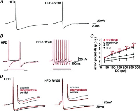
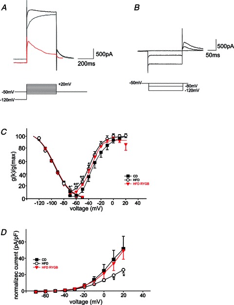
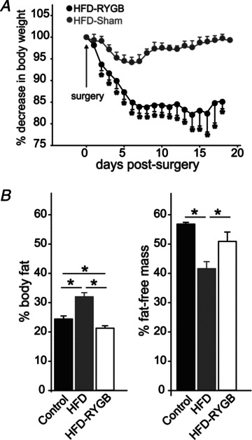
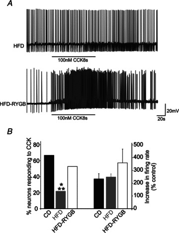
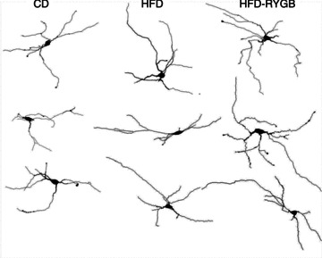
Diet-induced obesity (DIO) has been shown to alter the biophysical properties and pharmacological responsiveness of vagal afferent neurones and fibres, although the effects of DIO on central vagal neurones or vagal efferent functions have never been investigated. The aims of this study were to investigate whether high-fat diet-induced DIO also affects the properties of vagal efferent motoneurones, and to investigate whether these effects were reversed following weight loss induced by Roux-en-Y gastric bypass (RYGB) surgery. Whole-cell patch-clamp recordings were made from rat dorsal motor nucleus of the vagus (DMV) neurones in thin brainstem slices. The DMV neurones from rats exposed to high-fat diet for 12-14 weeks were less excitable, with a decreased membrane input resistance and decreased ability to fire action potentials in response to direct current pulse injection. The DMV neurones were also less responsive to superfusion with the satiety neuropeptides cholecystokinin and glucagon-like peptide 1. Roux-en-Y gastric bypass reversed all of these DIO-induced effects. Diet-induced obesity also affected the morphological properties of DMV neurones, increasing their size and dendritic arborization; RYGB did not reverse these morphological alterations. Remarkably, independent of diet, RYGB also reversed age-related changes of membrane properties and occurrence of charybdotoxin-sensitive (BK) calcium-dependent potassium current. These results demonstrate that DIO also affects the properties of central autonomic neurones by decreasing the membrane excitability and pharmacological responsiveness of central vagal motoneurones and that these changes were reversed following RYGB. In contrast, DIO-induced changes in morphological properties of DMV neurones were not reversed following gastric bypass surgery, suggesting that they may be due to diet, rather than obesity. These findings represent the first direct evidence for the plausible effect of RYGB to improve vagal neuronal health in the brain by reversing some effects of chronic high-fat diet as well as ageing. Vagovagal neurocircuits appear to remain open to modulation and adaptation throughout life, and understanding of these mechanisms may help in development of novel interventions to alleviate environmental (e.g. dietary) ailments and also alter neuronal ageing.


Figures

Figure 2
Figure 2. Gastic bypass surgery increases the action potential firing of neurones in the dorsal motor nucleus of the vagus (DMV)
A, representative traces illustrating the effects of HFD and RYGB on action potential properties. The DMV neurones were current clamped at −60 mV prior to injection of a short (15 ms) depolarizing current pulse sufficient to evoke the firing of a single action potential at current pulse offset. Note that the amplitude and duration of the action potential after-hyperpolarization was decreased significantly following RYGB. B, HFD and HFD-RYGB neurones were current clamped at −60 mV prior to injection of long (400 ms) depolarizing current pulses of increasing magnitude (from 30 to 270 pA). Note that, following RYGB, DMV neurones fired a greater number of action potentials. C, graphical representation of the frequency of action potential firing (expressed as pulses per second, p.p.s.) in DMV neurones from juvenile-CD, CD, HFD and HFD-RYGB rats. Note that, at all frequencies of current injection, CD DMV neurones were less excitable and fired fewer action potentials than juvenile-CD DMV neurones. The HFD further decreased DMV neurone excitability at all frequencies of current injection. Roux-en-Y gastric bypass reversed the effects of ageing and diet-induced obesity on DMV neuronal excitability. *P < 0.05 vs. control; **P < 0.05 vs. HFD. D, representative traces illustrating the effects of apamin (100 nm) and charybdotoxin (40 nm) on the action potential after-hyperpolarization in HFD (left) and HFD-RYGB (right) DMV neurones. Note that, in HFD neurones, both apamin and charybdotoxin decreased the amplitude and duration of the after-hyperpolarization, suggesting the presence of both small conductance (SK) and large conductance (BK) calcium-dependent potassium currents. In contrast, following RYGB, only the apamin-sensitive small conductance (SK) calcium-dependent potassium current was observed, and charybdotoxin had no effect on after-hyperpolarization amplitude or duration.
Figure 3
Figure 3. Gastric bypass surgery alters voltage-dependent potassium currents in DMV neurones
A, representative traces illustrating the activation of the ensemble outward potassium currents (upper trace, black), or IKV (middle trace, grey) upon stepping the neuronal membrane from −120 mV (IA) or −50 mV (IKV) to +20 mV (IA). The lower (red) trace is the current obtained by subtraction of IKV from the ensemble current. The peak current obtained consists principally of IA. B, the voltage dependence of IA inactivation was measured in DMV neurones voltage clamped at −50 mV. Neurones were then step hyperpolarized (400 ms duration) in 10 mV increments, to −120 mV before being repolarized to −50 mV. The resultant current values were normalized (Imax= 100) and averaged. For the purposes of clarity, only traces from −50, −80 and −120 mV are shown. C, graphical representation of the voltage dependence of IA inactivation and activation. Note that while the voltage dependence of IA inactivation was unchanged between CD, HFD and HFD-RYGB neurones, in contrast there was a leftward shift in the voltage dependence of IA activation in HFD and HFD-RYGB neurones, with half-maximal activations of −30, −40 and −35 mV for CD, HFD and HFD-RYGB neurones, respectively. *P < 0.05 CD vs. HFD; #P < 0.05 CD vs. HFD-RYGB; P < 0.05 HFD vs. HFD-RYGB. D, graphical representation of the normalized current–voltage relationship of the delayed rectifier, IKV. Note that, at potentials positive to +10 mV, the magnitude of IKV was reduced in DIO neurones, and this reduction was reversed following RYGB. *P < 0.05 HFD vs. CD and HFD-RYGB neurones. A) Representative traces showing the activation of the ensemble of potassium and calcium currents (upper trace) or IKV (middle trace, red) upon stepping to +20 mV from −120 mV (ensemble currents) or from −50 mV (IKV). The lower trace is the current obtained upon subtraction of the IKV from the ensemble current. The peak current obtained comprises IA mainly. For the purposes of clarity, only traces from –120 and −50 mV are shown. Similar results were obtained upon perfusion of the ensemble currents with 5 mm 4-AP (not shown). B) Activation curve of IKV in DMV neurones from untreated (control) and perivagal-CAP treated (capsaicin) rats. Note that at potentials positive to 0 mV, the peak IKV from capsaicin neurones is significantly reduced compared with control. C) Representative traces showing the inactivation of IA obtained upon hyperpolarization of the membrane from −50 to −120 mV and repolarization to −50 mV. For clarity purposes, only traces from −50, −80 and −120 mV are shown.
Figure 1
Figure 1. Graphical representation of the weight loss induced by gastric bypass surgery
A, rats were fed a high-fat diet (HFD; 60% kcal from fat) for 12 weeks prior to gastric bypass surgery (RYGB) or sham surgery. Rat weights were monitored daily from the day of surgery until experimentation. Note that, despite being maintained on the same HFD postsurgery, RYGB rats lost an average of 19.5 ± 1.6% body weight, while the weight of sham-operated rats was unchanged (0.5 ± 0.6% starting body weight). B, graphical representation of the 1H NMR measurements of body composition in control diet-fed (CD) rats (n= 5), HFD rats (n= 23) and HFD-RYGB rats (n= 8). Note that HFD-induced obesity significantly increased fat mass and decreased fat free mass, relative to CD rats, and these changes were reversed following RYGB. *P < 0.05.
Figure 4
Figure 4. Gastric bypass surgery restores the responsiveness of DMV neurones to CCK
A, representative traces from HFD (top trace) and HFD-RYGB DMV neurones (bottom trace) illustrating the response to superfusion with CCK (100 nm). Neurones were current clamped to allow a spontaneous action potential firing frequency of ∼1 Hz, before superfusion with 100 nm CCK for a period of time sufficient for the response to reach a plateau. The majority of HFD neurones were unresponsive to CCK, whereas, following RYGB, the majority of DMV neurones responded to CCK with a membrane depolarization and an increase in action potential firing. B, graphical representation of the proportion of CD, HFD and HFD-RYGB neurones that responded to CCK with an increase in action potential firing rate (left bars) and the magnitude of the increase in firing frequency (right bars). *P < 0.05 vs. control; **P < 0.05 vs. RYGB.
Figure 5
Figure 5. Gastric bypass surgery does not alter the morphological properties of DMV neurones
Computer-aided reconstructions of representative Neurobiotin-filled neurones from CD, HFD and HFD-RYGB rats. Note that, compared with neurones from control rats, diet-induced obesity increased the extent of dendritic arborization. These morphological changes were not reversed by RYGB, suggesting that they are due to the diet, rather than obesity.

References

    1. Abizaid A, Horvath TL. Brain circuits regulating energy homeostasis. Regul Pept. 2008;149:3–10. - PMC - PubMed
    1. Alywin ML, Horowitz JM, Bonham AC. NMDA receptors contribute to primary visceral afferent transmission in the nucleus of the solitary tract. J Neurophysiol. 1997;77:2539–2548. - PubMed
    1. Andresen MC, Yang M. Non-NMDA receptors mediate sensory afferent synaptic transmission in medial nucleus tractus solitarius. Am J Physiol Heart Circ Physiol. 1990;259:H1307–H1311. - PubMed
    1. Appleyard SM, Bailey TW, Doyle MW, Jin YH, Smart JL, Low MJ, Andresen MC. Proopiomelanocortin neurons in nucleus tractus solitarius are activated by visceral afferents: regulation by cholecystokinin and opioids. J Neurosci. 2005;25:3578–3585. - PMC - PubMed
    1. Babic T, Browning KN, Travagli RA. Differential organization of excitatory and inhibitory synapses within the rat dorsal vagal complex. Am J Physiol Gastrointest Liver Physiol. 2011;300:G21–G32. - PMC - PubMed

Publication types

Substances