Review
. 2013 Jul 1;591(13):3167-78.
doi: 10.1113/jphysiol.2012.249482. Epub 2013 Mar 4.

Single Ca2+ channels and exocytosis at sensory synapses

Affiliations
Review

Single Ca2+ channels and exocytosis at sensory synapses

Mean-Hwan Kim et al. J Physiol. .

Abstract

Hair cell synapses in the ear and photoreceptor synapses in the eye are the first synapses in the auditory and visual system. These specialized synapses transmit a large amount of sensory information in a fast and efficient manner. Moreover, both small and large signals with widely variable kinetics must be quickly encoded and reliably transmitted to allow an animal to rapidly monitor and react to its environment. Here we briefly review some aspects of these primary synapses, which are characterized by a synaptic ribbon in their active zones of transmitter release. We propose that these synapses are themselves highly specialized for the task at hand. Photoreceptor and bipolar cell ribbon synapses in the retina appear to have versatile properties that permit both tonic and phasic transmitter release. This allows them to transmit changes of both luminance and contrast within a visual field at different ambient light levels. By contrast, hair cell ribbon synapses are specialized for a highly synchronous form of multivesicular release that may be critical for phase locking to low-frequency sound-evoked signals at both low and high sound intensities. The microarchitecture of a hair cell synapse may be such that the opening of a single Ca(2+) channel evokes the simultaneous exocytosis of multiple synaptic vesicles. Thus, the differing demands of sensory encoding in the eye and ear generate diverse designs and capabilities for their ribbon synapses.


Figures

Figure 1
Figure 1. Multiquantal EPSCs triggered by the voltage-dependent opening of Ca2+ channels in hair cells
A, paired recordings between a presynaptic bullfrog auditory hair cell and its afferent fibre. The hair cell was dialysed with 2 mm EGTA and held at −90 mV. The hair cell was then depolarized to various membrane potentials in 3 mV increments (shown in red). At −72 mV, only small-amplitude EPSCs are observed (presumably single vesicle or quanta events). However, a further small depolarization to −69 mV (at which Ca2+ current starts to activate) suddenly evokes large-amplitude EPSCs together with small amplitude events (marked with a blue asterisk). The large EPSC events (presumably multiquantal) become more frequent with further depolarization to −66 mV. These events occur in a stochastic manner and may reflect the highly synchronous release of several vesicles (modified from Li et al. 2009). B, paired recordings from a hair cell dialysed with 10 mm BAPTA and held at −90 mV. In this recording depolarizations to −69 mV and −66 mV did not evoke large EPSCs, presumably because the large concentration of BAPTA, a fast Ca2+ buffer, reduced Ca2+ nanodomain concentrations. However, hair cell depolarization to −57 mV evoked large-amplitude EPSCs. The Ca2+ current at −57 mV is substantial and thus saturates the BAPTA at the active zone. The Ca2+ concentrations can then reach levels sufficient to evoke release. Note that large-amplitude EPSCs occur together with small amplitude EPSC events (modified from Li et al. 2009). C, unitary voltage-gated CaV1.3 L-type Ca2+ channel currents from mouse inner hair cells (modified from Zampini et al. 2010). Representative unitary currents from cell attached recordings. The membrane patch potentials (red) are shown next to the traces. The pipette solution contained 5 mm Ca2+ and BayK 8644, an L-type Ca2+ channel agonist. The grey lines indicate the closed channel state. At hyperpolarized potentials of −67 mV relatively few channel openings occur (open probability Po low) and the open time is short (about 0.5–1 ms), but the single Ca2+ channel current is large. Note that Ca2+ channels can open and close quickly (flickers). At the more depolarized potentials of −57 mV and −37 mV, more openings occur (higher Po) and open times are longer, but the single Ca2+ channel current is smaller due to reduced driving force. The very brief Ca2+ channel openings may trigger the release of single synaptic vesicles that produces small amplitude EPSCs, while the longer Ca2+ channel openings may trigger the highly synchronous release of several synaptic vesicles that produces large EPSC events.
Figure 3
Figure 3. Presynaptic gain control by Ca2+ channel coupling to the fusion sensor of synaptic vesicles at the active zone
A, for individual non-overlapping Ca2+ nanodomains the docked vesicles are tightly coupled with a single Ca2+ channel. Ca2+ influx through a single Ca2+ channel is able to evoke exocytosis reliably. In this case, the amount of release is linearly increased when the number of open Ca2+ channels is increased. Evoked release is insensitive to EGTA, a high-affinity but slow Ca2+ buffer. A small depolarization thus evokes a small number of open Ca2+ channels and reliable release. The synapse thus has high gain at low presynaptic membrane potentials (Vm). This is the modus operandi of mammalian rod bipolar cells for short depolarizing pulses (Jarsky et al. 2010) and for the squid giant synapse with presynaptic action potential depolarizations (Augustine et al. 1991). B, for microdomains individual docked vesicles are coupled to several presynaptic Ca2+ channels with overlapping Ca2+ domains. The overlapping Ca2+ domains cooperatively recruit vesicle exocytosis. In this case, the release rate is non-linearly increased when the number of open Ca2+ channels is increased. Release will increase in proportion to the third of fourth power of the increment in macroscopic Ca2+ current. Evoked release can be insensitive to EGTA. The synapse thus has low gain at small presynaptic Vm and high gain at high presynaptic Vm. This is the modus operandi of the squid giant synapse with long voltage-clamp depolarizing pulses (Augustine et al. 1991). C, a mixture of Ca2+ nanodomains and Ca2+ microdomains is also possible at some synapses. Some docked vesicles are tightly coupled with Ca2+ channels, while some docked vesicles are relatively far from a cluster of Ca2+ channels. This cluster of Ca2+ channels can produce a Ca2+ microdomain that can influence the release of distant vesicles. These Ca2+ microdomains can be reduced by EGTA. In this case the relationship between release and number of open Ca2+ channels can show both linear and non-linear components, and a pool of readily releasable synaptic vesicles will be insensitive to EGTA and a pool of vesicles will be sensitive to EGTA. This is perhaps the modus operandi of the Mb (mixed rod-cone) bipolar cell in goldfish retina (Mennerick & Matthews, 1996; Coggins & Zenisek, 2009). The dependence of release on presynaptic Vm may thus acquire a larger dynamic range, allowing the Mb bipolar cell terminal to transmit both rod and cone photoreceptor signals.
Figure 2
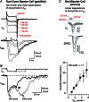
Figure 2. Non-linear and linear dependence of exocytosis on Ca2+ currents at retinal bipolar cell ribbon synapses
A, paired recording from an acutely dissociated goldfish Mb (mixed rod-cone) bipolar cell terminal and a cultured horizontal cell that expresses AMPA receptors. The horizontal cell is used to detect the glutamate release from the bipolar cell terminal. Superimposed responses were evoked by 10 ms pulses to +52 (in bold), −45, −43, −38 and −18 mV (from top to bottom) that elicited respectively larger calcium current (ICa) and glutamate-gated current (IGlu), except for the IGlu response to the +52 mV pulse (in bold), which occurred only during the repolarization from +52 mV to −68 mV, when a tail ICa is elicited (membrane potentials were corrected for a 8 mV liquid junction potential; von Gersdorff et al. 1998). Note that the synaptic delay (arrows) is considerable for weak depolarizations, which produce slow ICa activation and small ICa amplitudes. B, a dual-component IGlu is revealed by longer depolarizing pulses. Asynchronous release occurs after Ca2+ channel closure. Responses evoked by 50 ms and 100 ms pulses to −10 mV elicited, respectively, the bold and thin IGlu traces. The arrowhead marks the truncated second component of release evoked by the 50 ms pulse (von Gersdorff et al. 1998). A fast releasing pool of vesicles triggers the phasic first component of IGlu, whereas a delayed and slow second pool of released vesicles triggers a more sustained and tonic component of IGlu. C, paired recording from the mouse rod bipolar cell synapse. Exocytosis varies linearly with the number of open Ca2+ channels. Presynaptic voltage steps (1.5 ms) from −60 mV to potentials between −47 and −31 mV (in 4 mV increments; grey-scale from −47 mV (dark) to −31 mV (light)) elicit Ca2+ currents (top) and EPSCs (bottom). EPSC quantal content is plotted against QCa, illustrating a near-linear relationship between Ca2+ influx and exocytosis when the number of open presynaptic Ca2+ channels is altered. Data are normalized to the condition of the smallest voltage step in which 44% of responses are failures of transmission ([Ca2+]e= 1 mm; modified from Jarsky et al. 2010).
Figure 4
Figure 4. The spatial profile of a Ca2+ nanodomain
Left panel, a spherical synaptic ribbon is shown above an array of docked vesicles (red) and two clusters of Ca2+ channels (blue stripes). Right panel, the Ca2+ concentration underneath the synaptic ribbon in a bullfrog hair cell active zone of exocytosis after the opening of a single Ca2+ channel. The white circles show the locations of the docked spherical vesicles with a diameter of 30 nm. A Monte Carlo simulation was created to describe the diffusion of Ca2+ ions and the spatial extent of local Ca2+ nanodomains (Graydon et al. 2011). A 30 × 30 grid of 10 nm boxes was used to monitor free Ca2+ concentration at the presynaptic membrane surface over time while a single Ca2+ channel was opened. Shown are free Ca2+ concentrations averaged over 1 ms time bins beginning 5 ms after channel opening. The hair cell contained 2 mm EGTA as a mobile Ca2+ buffer. The single open channel current i=−0.54 pA at Vm=−70 mV. This current resulted in a widespread, large free Ca2+ nanodomain that peaked at about 300 μm near the open mouth of the Ca2+ channel. The presence of the docked vesicles and the ribbon as diffusion barriers and restrictors of available space led to an enhancement in Ca2+ concentration of tens of additional micromolars for tens of square nanometres (see Graydon et al. 2011). The simulations suggest that docked vesicles and the synaptic ribbon effectively ‘trap’ Ca2+ ions in a small and crowded volume. The synaptic ribbon may thus function as an aggregator of Ca2+ channels and docked vesicles, and if it is impermeable to Ca2+ ions, as a booster of Ca2+ nanodomain concentrations.

Similar articles

Cited by

References

    1. Augustine GJ, Adler EM, Charlton MP. The calcium signal for transmitter secretion from presynaptic nerve terminals. Ann N Y Acad Sci. 1991;635:365–381. - PubMed
    1. Babai N, Bartoletti TM, Thoreson WB. Calcium regulates vesicle replenishment at the cone ribbon synapse. J Neurosci. 2010;30:15866–15877. - PMC - PubMed
    1. Bartoletti TM, Jackman SL, Babai N, Mercer AJ, Kramer RH, Thoreson WB. Release from the cone ribbon synapse under bright light conditions can be controlled by the opening of only a few Ca2+ channels. J Neurophysiol. 2011;106:2922–2935. - PMC - PubMed
    1. Beaumont V, Llobet A, Lagnado L. Expansion of calcium microdomains regulates fast exocytosis at a ribbon synapse. Proc Natl Acad Sci U S A. 2005;102:10700–10705. - PMC - PubMed
    1. Beutner D, Voets T, Neher E, Moser T. Calcium dependence of exocytosis and endocytosis at the cochlear inner hair cell afferent synapse. Neuron. 2001;29:681–690. - PubMed

Publication types

LinkOut - more resources