

Review
doi: 10.1007/s12105-013-0430-7.
Epub 2013 Mar 5.
Fibro-osseous lesions of the maxillofacial bones
Affiliations
- PMID: 23459840
- PMCID: PMC3597154
- DOI: 10.1007/s12105-013-0430-7
Item in Clipboard
Review
Fibro-osseous lesions of the maxillofacial bones
Head Neck Pathol.
2013 Mar.
Abstract
Fibro-osseous lesions of the maxillofacial bones should be classified based on their radiographic growth pattern. This method can simplify this category of lesions, which have considerable overlapping histologic features. These neoplasms can be grouped into three categories: (a) fibrous dysplasia; (b) ossifying fibroma; (c) and osseous dysplasia. Important lesions in the differential diagnosis are osteoblastoma and giant cell reparative granuloma.
Figures
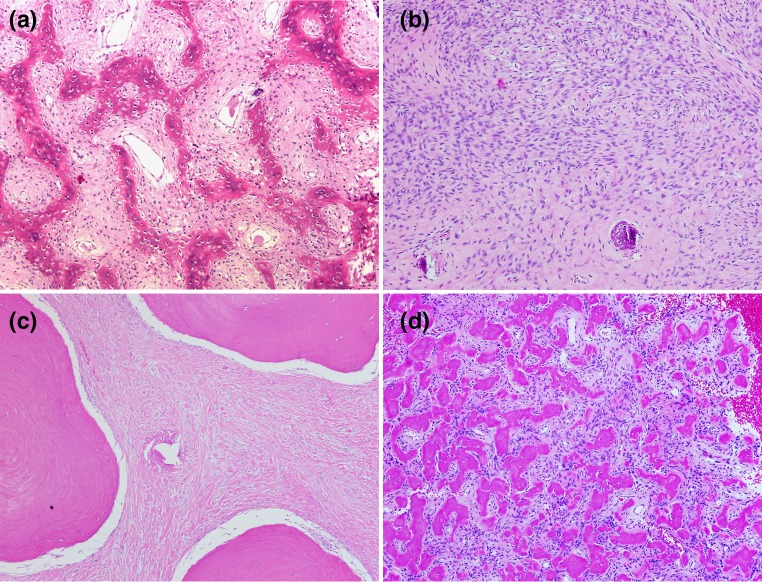
a A fibro-osseous pattern showing woven bone in the pattern of “Chinese letters” arranged in a spindle cell background (×40). b Fibro-osseous pattern with a predominance of spindle cells. Bone formation is minimal. c Fibro-osseous pattern showing broad plates of osteoid in a collagenized fibroblastic stroma. d Fibro-osseous pattern of globules of osteoid in a loose fibrous tissue background. This globular pattern has been called “cementum” (×40)
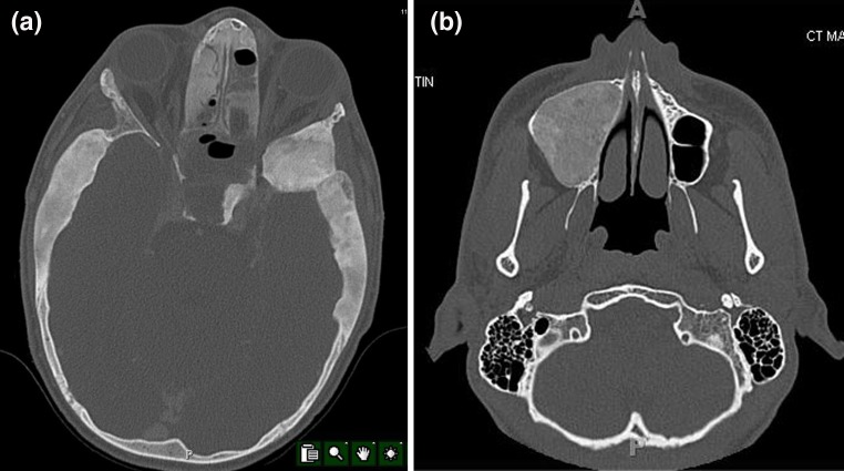
a A fibrous dysplasia of the skull and sinuses. There is diffuse thickening of broad areas of the calvarium as well as an intrasinus lesion. The thickening is poorly-circumscribed and has a “ground glass” appearance. b Ossifying fibroma of the maxillary sinus. The lesion has a “ground glass” appearance but it is extremely well-demarcated. No other lesions in the face and skull are present
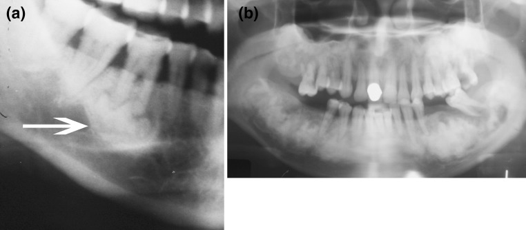
a Focal osseous dysplasia of the mandible. There is a periapical lucency filled with an irregular radiodensity (arrow). b Florid osseous dysplasia involving the apices of most of the teeth and the mandible. There is also involvement of the maxilla. The lesions are areas of radiolucency with central areas of irregular radiodensities
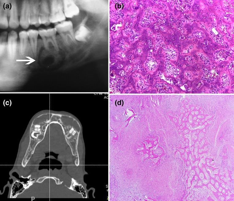
a Osteoblastoma of the mandible. There is a poorly-defined radiolucency with interlesional radiodensities in the mandible. This lesion may be mistaken radiographically for osseous dysplasia. b Osteoblastoma. There are interlacing, lace-like, seams of osteoid in a background of loose fibrovascular tissue. Osteoblasts are prominent which indicate that this is an osteoblastic neoplasm. c Multiple giant cell reparative granulomas in the mandible known as cherubism. There is extreme symmetrical expansion of the mandible. d A giant cell reparative granuloma in the healing phase. There is a zonal deposition of reactive bone surrounding central areas of fibrous tissue. The giant cells are no longer present in this stage. This lesion may be mistaken for a fibro-osseous lesion
References
-
- Sissons HA, Steiner GC, Dorfman HD. Calcified spherules in fibro-osseous lesions of bone. Arch Pathol Lab Med. 1993;117:284–290. - PubMed
-
- Tolman KG, Jubiz W, Sannella JJ, Madsen JA, Belsey RE, Goldsmith RS, Freston JW. Osteomalacia associated with anticonvulsant drug therapy in a pediatric outpatient population. Pediatrics. 1975;56:52. - PubMed
Publication types
MeSH terms
LinkOut - more resources
Full Text Sources
Other Literature Sources
Medical
