. 2013 May;81(5):1585-95.
doi: 10.1128/IAI.01117-12. Epub 2013 Mar 4.

The Plasmodium vivax merozoite surface protein 1 paralog is a novel erythrocyte-binding ligand of P. vivax

Affiliations

The Plasmodium vivax merozoite surface protein 1 paralog is a novel erythrocyte-binding ligand of P. vivax

Yang Cheng et al. Infect Immun. 2013 May.

Abstract

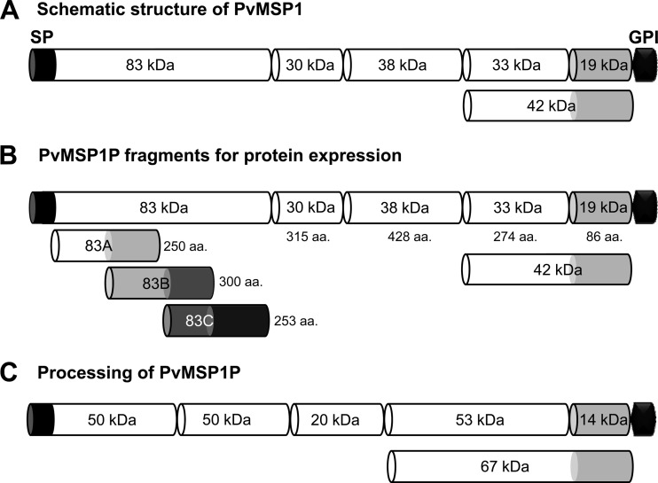
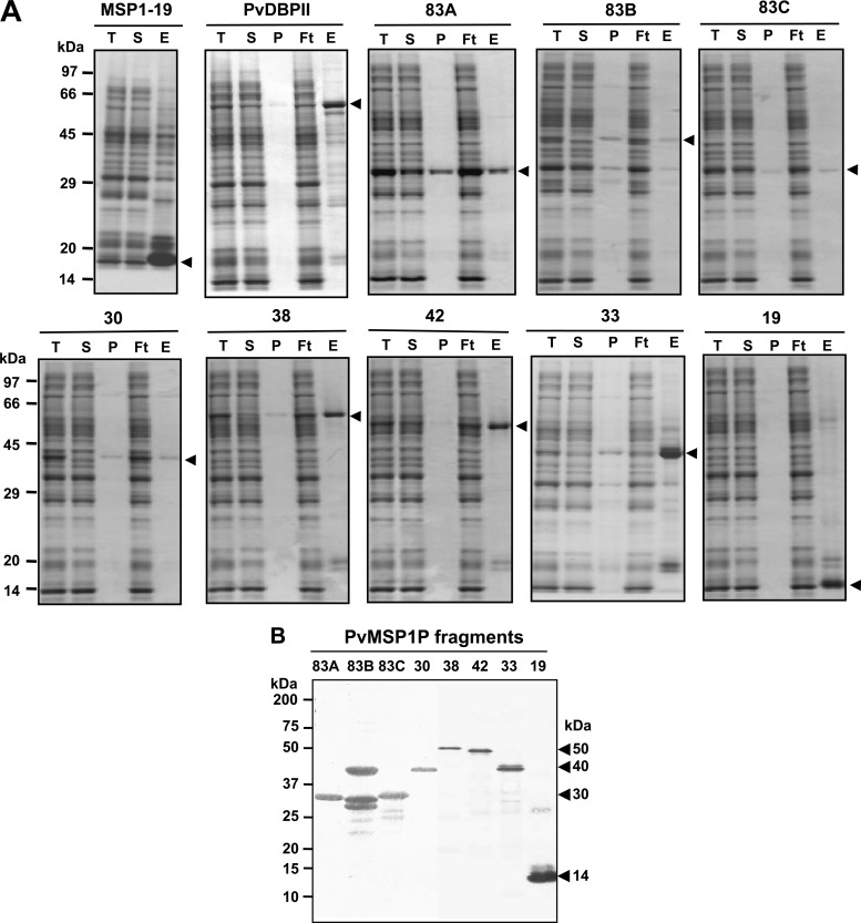
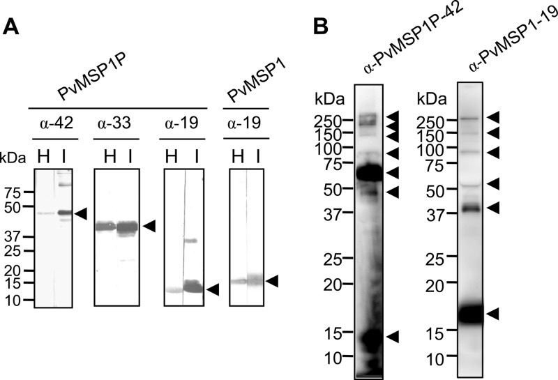
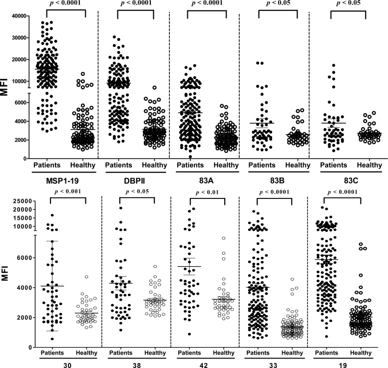
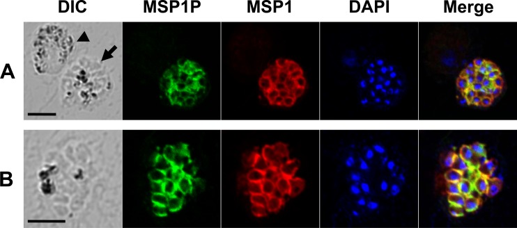
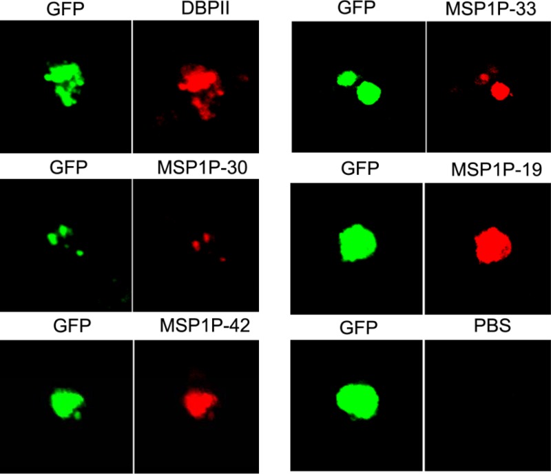
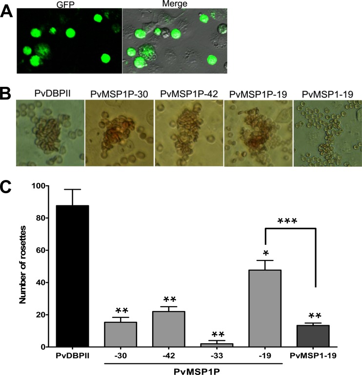
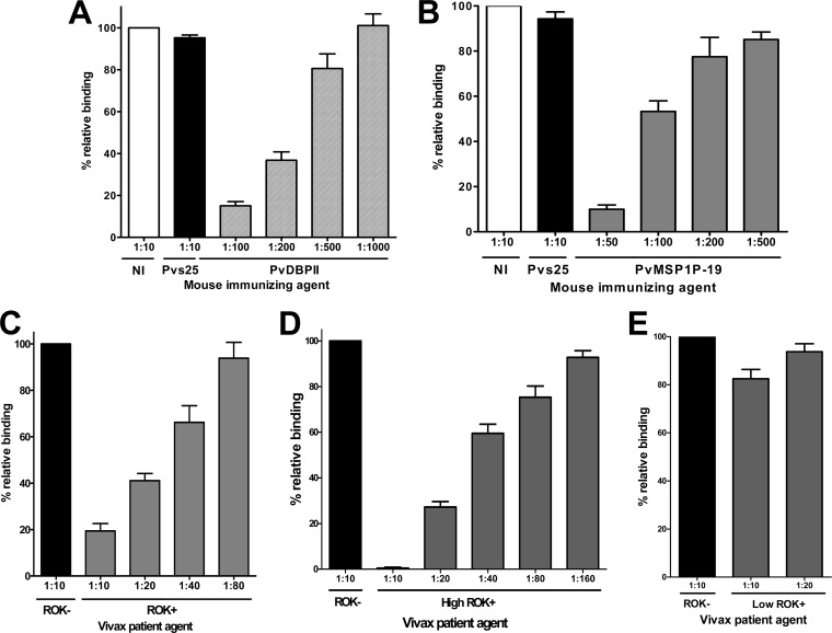
Merozoite surface protein 1 of Plasmodium vivax (PvMSP1), a glycosylphosphatidylinositol-anchored protein (GPI-AP), is a malaria vaccine candidate for P. vivax. The paralog of PvMSP1, named P. vivax merozoite surface protein 1 paralog (PvMSP1P; PlasmoDB PVX_099975), was recently identified and predicted as a GPI-AP. The similarities in genetic structural characteristics between PvMSP1 and PvMSP1P (e.g., size of open reading frames, two epidermal growth factor-like domains, and GPI anchor motif in the C terminus) led us to study this protein. In the present study, different regions of the PvMSP1P protein, demarcated based on the processed forms of PvMSP1, were expressed successfully as recombinant proteins [i.e., 83 (A, B, and C), 30, 38, 42, 33, and 19 fragments]. We studied the naturally acquired immune response against each fragment of recombinant PvMSP1P and the potential ability of each fragment to bind erythrocytes. The N-terminal fragment (83A) and two C-terminal fragments (33 and 19) reacted strongly with sera from P. vivax-infected patients, with 50 to 68% sensitivity and 95 to 96% specificity, respectively. Due to colocalization of PvMSP1P with PvMSP1, we supposed that PvMSP1P plays a similar role as PvMSP1 during erythrocyte invasion. An in vitro cytoadherence assay showed that PvMSP1P, especially the 19-kDa C-terminal region, could bind to erythrocytes. We also found that human sera from populations naturally exposed to vivax malaria and antisera obtained by immunization using the recombinant molecule PvMSP1P-19 inhibited in vitro binding of human erythrocytes to PvMSP1P-19. These results provide further evidence that the PvMSP1P might be an essential parasite adhesion molecule in the P. vivax merozoite and is a potential vaccine candidate against P. vivax.


Figures

Fig 1
Fig 1
Schematic diagram of PvMSP1, fragments of PvMSP1P for recombinant protein expression, and processing of native PvMSP1P. (A) Schematic diagram of processing of PvMSP1. (B) Demarcation of PvMSP1P, similar to the processed forms of PvMSP1, for protein expression of 8 fragments of PvMSP1P. For the N-terminal 83-kDa region, three 50-amino-acid overlapping fragments were expressed as 83A, 83B, and 83C. The MSP1P protein comprises 1,856 amino acids, with a calculated molecular mass of 214.5 kDa. The predicted signal peptide (SP; aa positions 1 to 28) and GPI anchor signal (GPI; aa 1835–1854) are shown. Eight fragments of MSP1P (83A [aa 29 to 278], 83B [aa 229 to 528], 83C [aa 479 to 731], 30 [aa 732 to 1046], 38 [aa 1047 to 1474], 42 [aa 1475 to 1834], 33 [aa 1475 to 1748], and 19 [aa 1749 to 1834]) were expressed as recombinant proteins. PvMSP1P-30, -42, -33, and -19 were used to raise specific antisera. (C) Hypothetical processing of PvMSP1P was predicted from Western blot results of the parasite lysate probed with anti-PvMSP1P-42 serum (Fig. 3B). aa, amino acid; kDa, kilodalton; SP, signal peptide; GPI, glycophosphatidylinositol anchor signal.
Fig 2
Fig 2
Recombinant protein expression and purification. (A) PvMSP1-19, PvDBPII, and all PvMSP1P fragments were synthesized using the wheat germ cell-free protein expression system and then purified with a Ni-Sepharose column. The purified MSP1-19 (15 kDa), DBPII (75 kDa), 83A (30 kDa), 83B (40 kDa), 83C (30 kDa), 30 (40 kDa), 38 (50 kDa), 42 (50 kDa), 33 (40 kDa), and 19 (14 kDa) fragments of PvMSP1P existed in soluble elution fractions. Arrowheads indicate specific bands for each recombinant protein. T, total translation mix; S, supernatant; P, pellet; Ft, flowthrough; E, elution. (B) The purified 83A, 83B, 83C, 30, 38, 42, 33, and 19 fragments of PvMSP1P were resolved by SDS-PAGE, transferred to a PVDF membrane, and probed with anti-His-tagged antibody. Arrowheads indicate specific bands for each recombinant protein.
Fig 3
Fig 3
Western blot analysis of recombinant PvMSP1P C-terminal fragments, PvMSP1-19, and schizont lysates probed with PvMSP1P and PvMSP1 immune sera. (A) Three C-terminal fragments of PvMSP1P (42, 33, and 19 kDa) and PvMSP1-19 were probed with the respective mouse immune sera (I) and with anti-His antibody (H). (B) Western blot analysis. Recognition of the native PvMSP1P and PvMSP1 antigen in the parasite lysate with mouse antisera against PvMSP1P-42 and rabbit antisera against PvMSP1-19, respectively, under reducing conditions. Arrowheads indicate the target bands and putative processed fragments.
Fig 4
Fig 4
IgG antibody responses to PvMSP1-19, PvDBPII, and eight fragments of PvMSP1P using protein microarrays. Immunoreactivity against each antigen with the sera of malaria patients (patients) and healthy individual samples (healthy) from ROK was determined. There were high specificity and significant differences in the total IgG prevalence for the eight PvMSP1P fragments between vivax patients and healthy individuals (P < 0.05). The P values were calculated using Student's t test. The bar indicates the mean ± standard deviation. MFI, mean fluorescence intensity.
Fig 5
Fig 5
Subcellular localization of PvMSP1P protein in asexual blood-stage parasites of P. vivax. (A) The acetone-fixed mature schizont (arrow) and a gametocyte (arrowhead) of P. vivax were dually labeled with mouse immune sera against PvMSP1P-42 (green) and rabbit immune sera against PvMSP1-19 (surface marker) (red). Nuclei are visualized with DAPI (blue). (B) Another mature schizont of P. vivax was also probed with the same antibodies as above. Bar represents 5 μm.
Fig 6
Fig 6
Transfection induced expression of parasite proteins on the surface of COS-7 cells. Expression of PvDBPII and PvMSP1P C-terminal fragments on the surface of COS-7 cells transfected with pEGFP-HSVgD1_PvDBPII and PvMSP1P-30, -42, -33, and -19 plasmid DNA was detected by IFA. Green fluorescent protein (GFP) (green, control), Alexa Fluor 543-conjugated goat anti-mouse antibody (red, MSP1P fragments), or Alexa Fluor 568-conjugated goat anti-rabbit antibody (red, DBPII) was used and visualized by confocal microscopy. Mouse antiserum against PBS was used as a negative control.
Fig 7
Fig 7
Binding of PvMSP1P fragments expressed on COS-7 cells to erythrocytes. (A) The transfection efficiency of each plasmid construct was calculated by counting green fluorescent cells (GFP) and COS-7 cells in bright field image (Merge). Average transfection efficiency was more than 80%. (B) Erythrocyte-binding rosettes formed on the surfaces of COS-7 cells expressing PvDBPII or different fragments of PvMSP1P or PvMSP1-19 were visualized under light microscopy. (C) The number of rosettes formed by the COS-7 cells transfected with genes coding for either PvDBPII or different fragments of PvMSP1P or PvMSP1-19. Detection of the transfection efficiency into COS-7 cells of all constructs by counting green signal cells within 30 microscope fields (×200). A positive result was defined as more than half the surface of the transfected cells covered with attached erythrocytes, and the total number of COS-7 cells per coverslip was recorded. Data are shown as the mean number of rosettes of three independent experiments, and the error bar represents ± standard deviation. Statistical differences between PvDBPII and the other proteins are indicated with a single asterisk (P < 0.05) and double asterisks (P < 0.001). Statistical difference between PvMSP1P-19 and PvMSP1-19 is shown with triple asterisks (P < 0.001).
Fig 8
Fig 8
Inhibition of erythrocyte binding to P. vivax MSP1P-19 expressed on COS-7 cells by naturally acquired antibodies and mouse anti-PvMSP1P-19 antibody. (A) COS-7 cells were transfected with pEGFP-HSVgD1_DBPII plasmid DNA expressing a GFP-DBPII fusion protein. Cells were subsequently incubated with anti-PvDBPII immune mouse sera at various dilutions before the addition of human erythrocytes. Binding was scored by counting the number of rosettes bound to COS-7 cells in 30 microscope fields (×200). Mouse antiserum against PBS as nonimmunized control (NI) and anti-Pvs25 (no erythrocyte-binding activity) immune mouse sera diluted 1:10 were included as negative controls (Pvs25). (B) Inhibition of erythrocyte binding to PvMSP1P-19 expressed on COS-7 cells by PvMSP1P immune mouse sera. Cells were then incubated with PvMSP1P immune mouse sera at various dilutions before the addition of human erythrocytes. (C) Inhibition of erythrocyte binding to PvMSP1P-19 expressed on COS-7 cells by vivax immune human sera. Cells were incubated with various dilutions of pooled sera from vivax malaria patients of ROK (ROK+) before the addition of human erythrocytes. Erythrocyte-binding activity was scored by counting the number of rosettes in 30 fields at a magnification of ×200. The percent binding was determined relative to a 1:10 dilution of pooled sera from areas of nonendemicity of ROK (ROK) as a negative control. Cells were incubated with various dilutions of the high-responding (high ROK+) (D) and low-responding (low ROK+) (E) vivax malaria sera before the addition of human erythrocytes. Error bars represent ± standard deviations.

References

    1. Mueller I, Galinski MR, Baird JK, Carlton JM, Kochar DK, Alonso PL, del Portillo HA. 2009. Key gaps in the knowledge of Plasmodium vivax, a neglected human malaria parasite. Lancet Infect. Dis. 9:555–566 - PubMed
    1. Gaur D, Mayer DC, Miller LH. 2004. Parasite ligand-host receptor interactions during invasion of erythrocytes by Plasmodium merozoites. Int. J. Parasitol. 34:1413–1429 - PubMed
    1. Cowman AF, Crabb BS. 2006. Invasion of red blood cells by malaria parasites. Cell 124:755–766 - PubMed
    1. Miller LH, Mason SJ, Clyde DF, McGinniss MH. 1976. The resistance factor to Plasmodium vivax in blacks. The Duffy-blood-group genotype, FyFy. N. Engl. J. Med. 295:302–304 - PubMed
    1. Menard D, Barnadas C, Bouchier C, Henry-Halldin C, Gray LR, Ratsimbasoa A, Thonier V, Carod JF, Domarle O, Colin Y, Bertrand O, Picot J, King CL, Grimberg BT, Mercereau-Puijalon O, Zimmerman PA. 2010. Plasmodium vivax clinical malaria is commonly observed in Duffy-negative Malagasy people. Proc. Natl. Acad. Sci. U. S. A. 107:5967–5971 - PMC - PubMed

Publication types

MeSH terms

LinkOut - more resources