. 2013 Apr 19;288(16):11407-15.
doi: 10.1074/jbc.M112.436659. Epub 2013 Mar 4.

Impaired intestinal calcium absorption in protein 4.1R-deficient mice due to altered expression of plasma membrane calcium ATPase 1b (PMCA1b)

Affiliations

Impaired intestinal calcium absorption in protein 4.1R-deficient mice due to altered expression of plasma membrane calcium ATPase 1b (PMCA1b)

Congrong Liu et al. J Biol Chem. .

Abstract

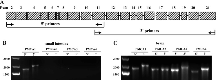
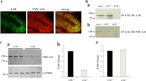
Protein 4.1R was first identified in the erythrocyte membrane skeleton. It is now known that the protein is expressed in a variety of epithelial cell lines and in the epithelia of many tissues, including the small intestine. However, the physiological function of 4.1R in the epithelial cells of the small intestine has not so far been explored. Here, we show that 4.1R knock-out mice exhibited a significantly impaired small intestinal calcium absorption that resulted in secondary hyperparathyroidism as evidenced by increased serum 1,25-(OH)2-vitamin D3 and parathyroid hormone levels, decreased serum calcium levels, hyperplasia of the parathyroid, and demineralization of the bones. 4.1R is located on the basolateral membrane of enterocytes, where it co-localizes with PMCA1b (plasma membrane calcium ATPase 1b). Expression of PMCA1b in enterocytes was decreased in 4.1(-/-) mice. 4.1R directly associated with PMCA1b, and the association involved the membrane-binding domain of 4.1R and the second intracellular loop and C terminus of PMCA1b. Our findings have enabled us to define a functional role for 4.1R in small intestinal calcium absorption through regulation of membrane expression of PMCA1b.


Figures

FIGURE 1.
FIGURE 1.
Expression and localization of protein 4.1R on small intestinal epithelia. A, Western blot analysis of 4.1R from the mucosa of the small intestine. Total cell lysates from 4.1R+/+ and 4.1R−/− duodenal and jejunal mucosae were probed with anti-4.1R exon 13 antibody. Note the detection of an ∼110-kDa band in 4.1R+/+ but not 4.1R−/− samples. B, immunolocalization of 4.1R on enterocytes. 4.1R+/+ and 4.1R−/− duodenal sections were stained with anti-4.1R exon 13 antibody. Note the staining of 4.1R on the basolateral membrane of enterocytes in 4.1R+/+ but not 4.1R−/− duodenum.
FIGURE 2.
FIGURE 2.
Altered small intestinal calcium absorption in 4.1R−/− mice. Calcium absorption was assessed by lavage assay as described under “Materials and Methods.” Note the significantly decreased uptake of calcium in 4.1R−/− mice (p < 0.01, n = 9).
FIGURE 3.
FIGURE 3.
Biochemical alterations in 4.1R knock-out mice. The levels of serum 1,25-(OH)2-vitamin D3 (1,25(OH)2D3; A), PTH (B), and calcium (C) from 3-, 6-, 12-, and 18-month-old 4.1R+/+ and 4.1R−/− mice were measured. Note the increased 1,25-(OH)2-vitamin D3 and PTH levels in all ages of 4.1R−/− mice, but the decreased level of serum calcium in 18-month-old mice (n = 9). *, p < 0.05.
FIGURE 4.
FIGURE 4.
Histological and pathological changes in 4.1R−/− parathyroid glands. A, representative hematoxylin/eosin staining of parathyroid glands from 4.1R+/+ and 4.1R−/− mice. Note the marked enlargement and hypercellularity of the 4.1R−/− parathyroid (20-fold) compared with its 4.1R+/+ counterpart. B, volumes of 4.1R+/+ and 4.1R−/− parathyroid glands. Data from 3-, 6-, 12-, and 18-month-old 4.1R+/+ and 4.1R−/− mice are plotted. *, p < 0.05. Note the increase in parathyroid gland volume at all ages.
FIGURE 5.
FIGURE 5.
Osteopenia in 4.1R−/− mice. A, representative x-ray images of the skeletons of 4.1R+/+ and 4.1R−/− mice. Images of vertebrae, hips, and femurs are shown. Note the decreased bone density in 4.1R−/− mice. Data are from a 1-year-old mouse. B, bone calcium content. Data from 3-, 6-, 12-, and 18-month-old 4.1R+/+ and 4.1R−/− mice are plotted. *, p < 0.05. Note the decreased calcium content in 4.1R−/− mice at all ages.
FIGURE 6.
FIGURE 6.
RT-PCR analysis of PMCA transcripts. A, schematic presentation of primer selection. B, amplification of PMCA transcripts from the small intestinal epithelium. The 5′- or 3′-portion of various PMCAs was amplified as described under “Materials and Methods” using cDNA from the small intestinal mucosa as a template. Note that only PMCA1b transcripts were amplified. C, amplification of PMCA transcripts from the brain. The 5′- or 3′-portion of various PMCAs was amplified as described for B using cDNA from the brain as a template. Note that all PMCA transcripts were amplified.
FIGURE 7.
FIGURE 7.
Association of 4.1R with PMCA1b on enterocytes. A, co-localization of 4.1R and PMCA1b. A section from a wild-type small intestine was co-stained with goat anti-4.1R exon 13 antibody (green) and rabbit anti-PMCA1b loop 2 antibody (red). Merged images show that 4.1R co-localized with PMCA1b on the bilateral membrane. B, co-immunoprecipitation of 4.1R with PMCA1b. The lysate from 4.1R+/+ or 4.1R−/− small intestinal mucosa was immunoprecipitated (IP) by anti-4.1R IgG and detected by anti-4.1R antibody (panel a) or anti-PMCA1b loop 2 antibody (panel b), respectively. IB, immunoblot. C, Western blot analysis. 20 μg of total protein from three 4.1R+/+ or three 4.1R−/− small intestinal mucosae was subjected to Western blotting using anti-PMCA1b loop 2 or anti-GAPDH antibody as shown in panel a, and the quantitative analysis is shown in panel b. D, quantitative real-time PCR, performed as described under “Materials and Methods.” No significant difference was detected between 4.1R+/+ and 4.1R−/− small intestines.
FIGURE 8.
FIGURE 8.
Direct binding between 4.1R and PMCA1b in vitro. A, schematic model of 4.1R structure. 4.1R contains four domains: the N-terminal 30-kDa membrane-binding domain, the 16-kDa domain, the internal 10-kDa spectrin-actin-binding domain, and the C-terminal 22/24-kDa domain. The 30-kDa domain consists of lobes A–C. B, schematic model of the PMCA1b structure. PMCA1b contains 10 membrane-spanning segments. Both the N and C termini are located on the cytosolic side of the membrane. In addition, a loop (designated loop 1 here) between transmembrane segments 2 and 3 and another large unit (designated loop 2 here) between transmembrane segments 4 and 5 are also facing the cytoplasmic side. C, binding of 4.1R to PMCA1b. Recombinant His-tagged 4.1R was incubated with GST-tagged fragments of PMCA1b. Binding was assayed by Western blotting using anti-His antibody for detection. Note the binding of 4.1R to loop 2 and the C terminus (C-ter) of PMCA1b. N-ter, N terminus of PMCA1b. D, binding of 4.1R domains to loop 2 and the C terminus of PMCA1b. His-tagged domains of 4.1R were incubated with the GST-tagged loop 2 or C terminus of PMCA1b. Binding was assayed as described for C. Note that only the 30-kDa domain bound to loop 2 and the C terminus of PMCA1b. E, binding of loop 2 or the C terminus of PMCA1b to subdomains of 30-kDa 4.1R. The GST-tagged loop 2 or C terminus of PMCA1b was incubated with maltose-binding protein-tagged lobes A–C of 30-kDa 4.1R. Binding was detected by Western blotting using anti-GST antibody. Note the binding of loop 2 to lobe A and the binding of C terminus to lobe C.

References

    1. Tchernia G., Mohandas N., Shohet S. (1981) Deficiency of skeletal membrane protein band 4.1 in homozygous hereditary elliptocytosis. Implications for erythrocyte membrane stability. J. Clin. Invest. 68, 454–460 - PMC - PubMed
    1. Salomao M., Zhang X., Yang Y., Lee S., Hartwig J. H., Chasis J. A., Mohandas N., An X. (2008) Protein 4.1R-dependent multiprotein complex: new insights into the structural organization of the red blood cell membrane. Proc. Natl. Acad. Sci. U.S.A. 105, 8026–8031 - PMC - PubMed
    1. Conboy J. G., Chan J., Mohandas N., Kan Y. W. (1988) Multiple protein 4.1 isoforms produced by alternative splicing in human erythroid cells. Proc. Natl. Acad. Sci. U.S.A. 85, 9062–9065 - PMC - PubMed
    1. Parra M. K., Gee S. L., Koury M. J., Mohandas N., Conboy J. G. (2003) Alternative 5′ exons and differential splicing regulate expression of protein 4.1R isoforms with distinct N termini. Blood 101, 4164–4171 - PubMed
    1. Gascard P., Lee G., Coulombel L., Auffray I., Lum M., Parra M., Conboy J. G., Mohandas N., Chasis J. A. (1998) Characterization of multiple isoforms of protein 4.1R expressed during erythroid terminal differentiation. Blood 92, 4404–4414 - PubMed

Publication types

MeSH terms

LinkOut - more resources