. 2013;8(2):e58052.
doi: 10.1371/journal.pone.0058052. Epub 2013 Feb 27.

PRAME is a golgi-targeted protein that associates with the Elongin BC complex and is upregulated by interferon-gamma and bacterial PAMPs

Affiliations

PRAME is a golgi-targeted protein that associates with the Elongin BC complex and is upregulated by interferon-gamma and bacterial PAMPs

Frances R Wadelin et al. PLoS One. 2013.

Erratum in

Abstract

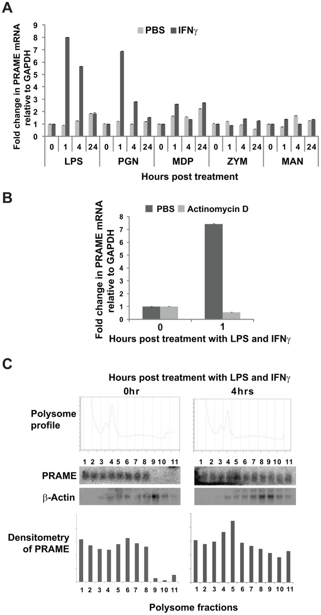
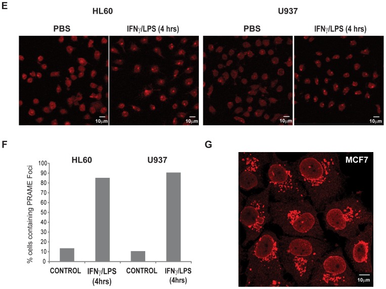
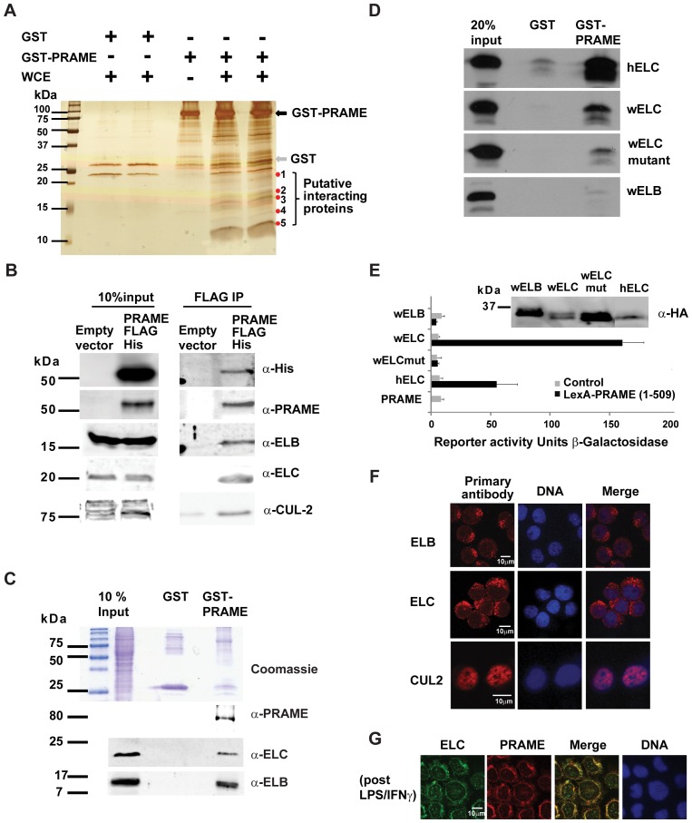
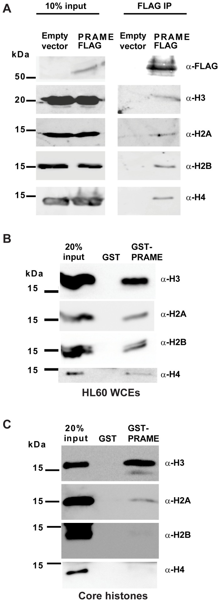
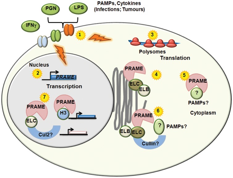
Preferentially expressed antigen in melanoma (PRAME) has been described as a cancer-testis antigen and is associated with leukaemias and solid tumours. Here we show that PRAME gene transcription in leukaemic cell lines is rapidly induced by exposure of cells to bacterial PAMPs (pathogen associated molecular patterns) in combination with type 2 interferon (IFNγ). Treatment of HL60 cells with lipopolysaccharide or peptidoglycan in combination with IFNγ resulted in a rapid and transient induction of PRAME transcription, and increased association of PRAME transcripts with polysomes. Moreover, treatment with PAMPs/IFNγ also modulated the subcellular localisation of PRAME proteins in HL60 and U937 cells, resulting in targeting of cytoplasmic PRAME to the Golgi. Affinity purification studies revealed that PRAME associates with Elongin B and Elongin C, components of Cullin E3 ubiquitin ligase complexes. This occurs via direct interaction of PRAME with Elongin C, and PRAME colocalises with Elongins in the Golgi after PAMP/IFNγ treatment. PRAME was also found to co-immunoprecipitate core histones, consistent with its partial localisation to the nucleus, and was found to bind directly to histone H3 in vitro. Thus, PRAME is upregulated by signalling pathways that are activated in response to infection/inflammation, and its product may have dual functions as a histone-binding protein, and in directing ubiquitylation of target proteins for processing in the Golgi.


Conflict of interest statement

Competing Interests: The authors have declared that no competing interests exist.

Figures

Figure 1
Figure 1. Transcriptional and translational regulation of PRAME by PAMPs/IFNγ.
(A) RT-qPCR measurements of PRAME gene expression relative to control GAPDH in HL60 cells in response to treatment with different PAMPs including lipopolysaccharide (LPS), peptidoglycan (PGN), muramyl dipeptide (MDP), zymosan (ZYM) and mannan (MAN) either alone (PBS) or in combination with IFNγ. Numbers on the x-axis indicate the time in hrs post-treatment. qPCR quantifications were performed in triplicate and the data shown represents the mean of two independent experiments, with error bars indicating standard deviations. The data is presented as fold induction relative to levels obtained at 0 hr (baseline). (B) RT-qPCR experiment performed as in (A) showing effect of pre-treatment with actinomycin D (10 μg/ml) or PBS on induction of PRAME transcript in HL60 cells by LPS/IFNγ (1 hour). (C) Association of PRAME transcripts with polysomes in HL60 cells following treatment with LPS/IFNγ for 0 and 4 hrs. Following cycloheximide treatment and sucrose density centrifugation of HL60 cell lysates, gradients were fractionated with continuous monitoring at 254 nm, to generate polysome profiles (top panel). RNA extracted from polysome fractions was analysed by Northern blotting and the PRAME transcripts visualised by phosphoimager (middle panels) and quantified by densitometry (bottom panels). β-actin was used as a control probe.
Figure 2
Figure 2. PRAME localises to the Golgi network following LPS/IFNγ treatment.
(A) HEK293 cells (upper panels) were transiently transfected with PRAME-EGFP (green) and stained with α-PRAME antibody (red) to confirm the identity of the overexpressed EGFP fusion protein. U2OS cells (lower panels) were cotranfected with GFP (green) and PRAME-FLAG (red). Merged images indicate the extent of coincidence of the EGFP and α-PRAME signals, and nuclear DNA is indicated (blue). The right hand panels are western blots showing detection of GFP or PRAME-EGFP proteins in whole cell extracts of transfected U2OS cells. (B) Immunostaining of endogenous PRAME in HL60 cells using α-PRAME antibody following treatment with PBS, LPS/IFNγ or PGN/IFNγ for 4 hrs. (C) Immunostaining of endogenous PRAME in U937 cells with α-PRAME following treatment with LPS/IFNγ for 0, 1 and 4 hrs. (D) HL60 cells treated with LPS/IFNγ for 4 hrs and immunostained with α-Golgi 58K (green) and α-PRAME (red). Merged images show the extent of colocalisation of both proteins. For immunofluorescence (A–D), nuclear DNA was stained using Hoechst 33258 and images were captured using a LSM510 confocal laser scanning microscope. (E) Immunostaining of endogenous PRAME in HL60 cells using α-PRAME antibody following treatment with PBS or LPS/IFNγ for 4 hrs. (F) Quantification (n = 60) of the percentage of cells in (E) containing PRAME cytoplasmic foci in treated cells or controls. (G) Immunostaining of endogenous PRAME in MCF-7 cells using α-PRAME antibody.
Figure 3
Figure 3. PRAME associates with the Elongin BC complex.
(A) SDS-PAGE and silver staining showing affinity capture of proteins from HL60 whole cell extracts (WCE) by immobilised GST or GST-PRAME proteins. GST and GST-PRAME proteins are indicated. Putative PRAME-specific bands are indicated and bands of approximately 12 kDa and 17 kDa were excised for mass spectrometry analysis. (B) Co-immunoprecipitation of PRAME with Elongin complex components. Whole cell extracts of HEK293 cells transfected with PRAME-FLAG-6xHis (or empty vector control) applied to anti-FLAG sepharose beads as described in Materials and Methods. After extensive washing, co-purified PRAME and E3 ubiquitin ligase complex components were detected by western blotting using specific antibodies as indicated. (C) GST-pulldown experiment showing binding of ELB and ELC proteins in HL60 whole cell extracts to GST or GST-PRAME proteins. The top panel is a Coomassie-stained gel showing the input whole cell extract, and the purified GST and GST-PRAME proteins. The lower panels are western blots revealing PRAME, ELB and ELC proteins bound to GST proteins. (D) GST-pulldown experiments revealing interactions of 35 [S]-labelled in vitro translated human ELC (hELC), C.elegans ELC (wELC), C.elegans ELC (L47D-L49D-Y88D-Y91D) (wELC mutant) and C.elegans ELB proteins with GST or GST-PRAME. (E) Yeast two hybrid assays of LexA-PRAME interactions with GAL4 AD-fused human ELC (hELC) or C.elegans proteins (wELB, wELC, wELC mutant). Western blots of the HA-tagged elongin fusion proteins are also shown. Reporter activity is expressed as β-galactosidase activity normalised to amount of protein in the extracts. (F) Immunofluorescence staining showing subcellular localisation of endogenous ELC, ELB and CUL2 proteins in HL60 cells. (G) Immunofluorescence staining showing colocalisation of endogenous ELC and PRAME proteins in HL60 cells following treatment with LPS/IFNγ for 4 hours.
Figure 4
Figure 4. Binding of PRAME to histone H3.
(A) Co-immunoprecipitation of endogenous histones with PRAME-FLAG-6xHis isolated from extracts of transfected HEK293 cells. Input lanes (left panels) show the presence of endogenous or FLAG-tagged proteins in extracts from cells transfected with PRAME-FLAG or empty vector. After IP with anti-FLAG antibody, immunoblots were performed with the antibodies indicated. (B) GST-pulldown experiment showing association of histones with GST or GST-PRAME proteins. Whole cell extracts of HL60 cells were incubated with immobilised GST or GST-PRAME proteins, and bound proteins separated by SDS-PAGE. Immunoblots were performed with specific antibodies to detect association of histones H2A, H2B, H3 and H4 with GST proteins. (C) Direct association of histones with GST or GST-PRAME proteins. Core histone preparations were incubated with GST beads. Following extensive washing, bound histones were separated by SDS-PAGE and revealed by western blotting using specific antibodies as indicated.
Figure 5
Figure 5. Potential Functions and Regulation of PRAME.
Activation of TLRs and other signalling receptors by PAMPs and cytokines associated with infections or tumours (1) results in both transcriptional (2) and translational (3) upregulation of PRAME. Cytoplasmic PRAME can associate with Elongin proteins located in the Golgi/ER network (4). Association of PRAME with intracellular PAMPs or other molecules (5) may facilitate their targeting to the Golgi for modification, secretion or destruction by Elongin/Cullin Ubiquitin ligases (6). Interactions of PRAME proteins with Elongins, Cul2 or Histone H3 in the nucleus (7) may be involved in the regulation of gene expression.

References

    1. Wadelin F, Fulton J, McEwan PA, Spriggs KA, Emsley J, et al. (2010) Leucine-rich repeat protein PRAME: expression, potential functions and clinical implications for leukaemia. Mol Cancer 9: 226. - PMC - PubMed
    1. Ikeda H, Lethe B, Lehmann F, van Baren N, Baurain JF, et al. (1997) Characterization of an antigen that is recognized on a melanoma showing partial HLA loss by CTL expressing an NK inhibitory receptor. Immunity 6: 199–208. - PubMed
    1. Atanackovic D, Luetkens T, Kloth B, Fuchs G, Cao Y, et al. (2011) Cancer-testis antigen expression and its epigenetic modulation in acute myeloid leukemia. Am J Hematol 86: 918–922. - PubMed
    1. Kessler JH, Beekman NJ, Bres-Vloemans SA, Verdijk P, van Veelen PA, et al. (2001) Efficient identification of novel HLA-A(*)0201-presented cytotoxic T lymphocyte epitopes in the widely expressed tumor antigen PRAME by proteasome-mediated digestion analysis. J Exp Med 193: 73–88. - PMC - PubMed
    1. Matsushita M, Ikeda H, Kizaki M, Okamoto S, Ogasawara M, et al. (2001) Quantitative monitoring of the PRAME gene for the detection of minimal residual disease in leukaemia. Br J Haematol 112: 916–926. - PubMed

Publication types

MeSH terms

LinkOut - more resources