. 2013;8(2):e58057.
doi: 10.1371/journal.pone.0058057. Epub 2013 Feb 27.

Connexin43 hemichannel-mediated regulation of connexin43

Affiliations

Connexin43 hemichannel-mediated regulation of connexin43

Kai Li et al. PLoS One. 2013.

Abstract

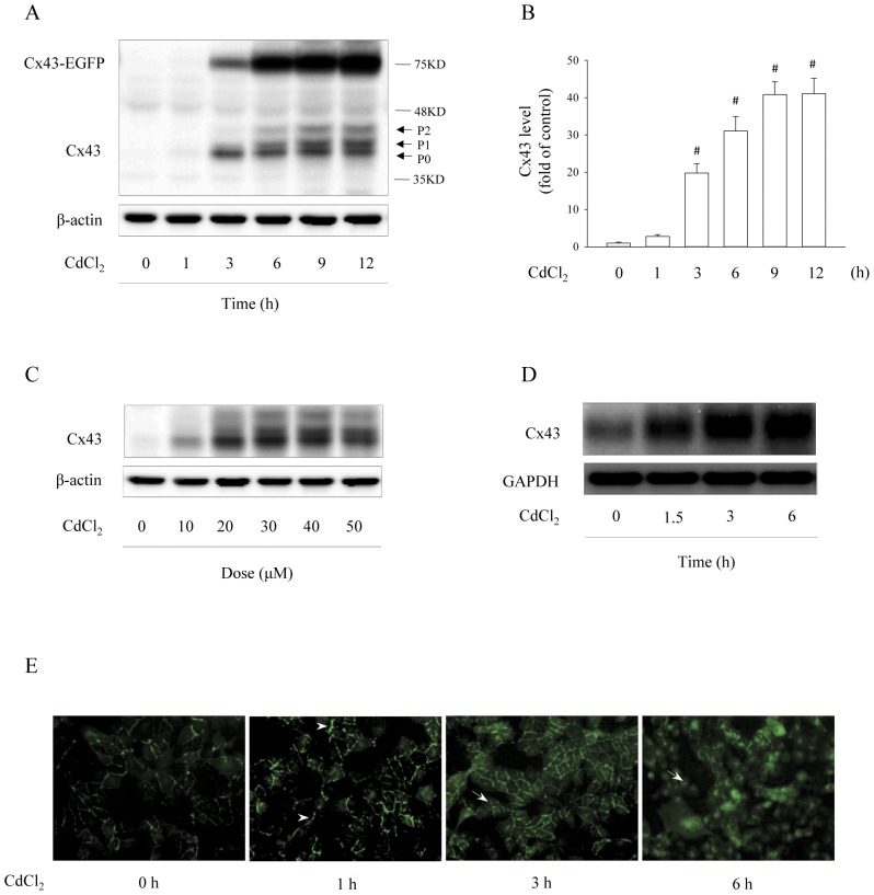
Background: Many signaling molecules and pathways that regulate gap junctions (GJs) protein expression and function are, in fact, also controlled by GJs. We, therefore, speculated an existence of the GJ channel-mediated self-regulation of GJs. Using a cell culture model in which nonjunctional connexin43 (Cx43) hemichannels were activated by cadmium (Cd(2+)), we tested this hypothesis.

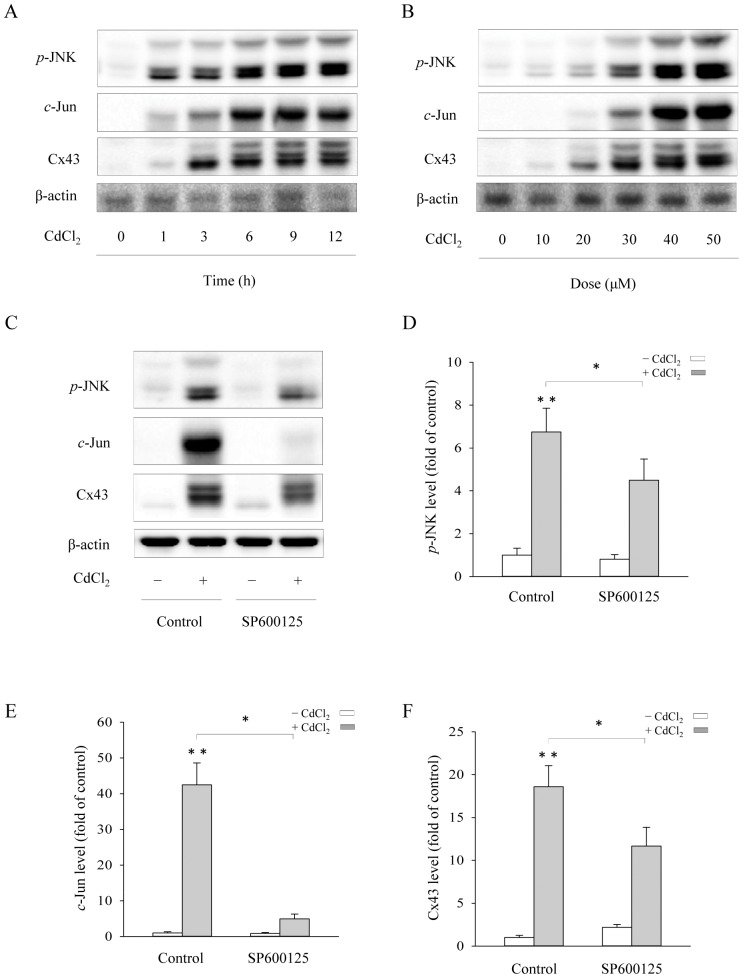
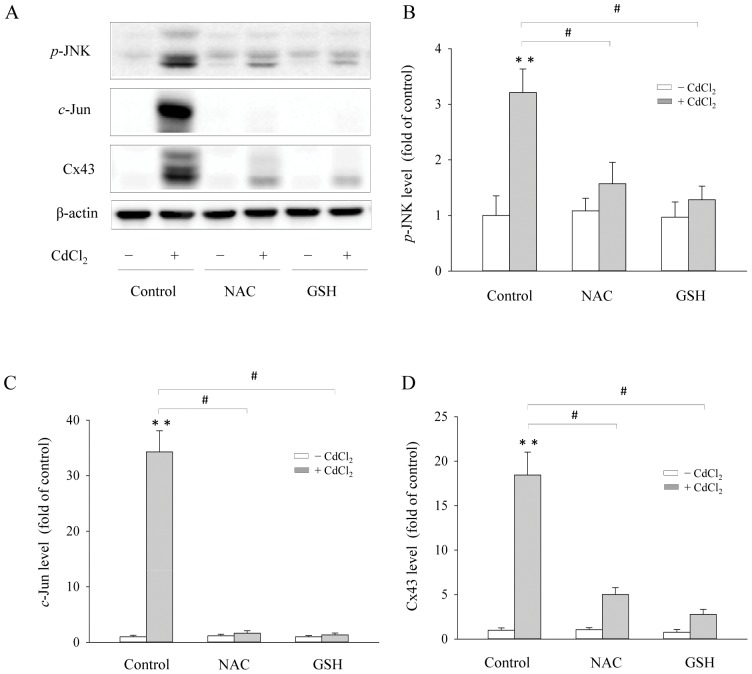
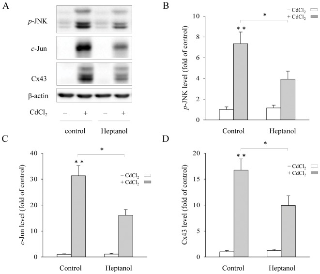
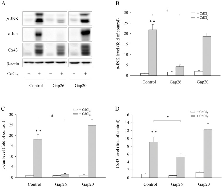
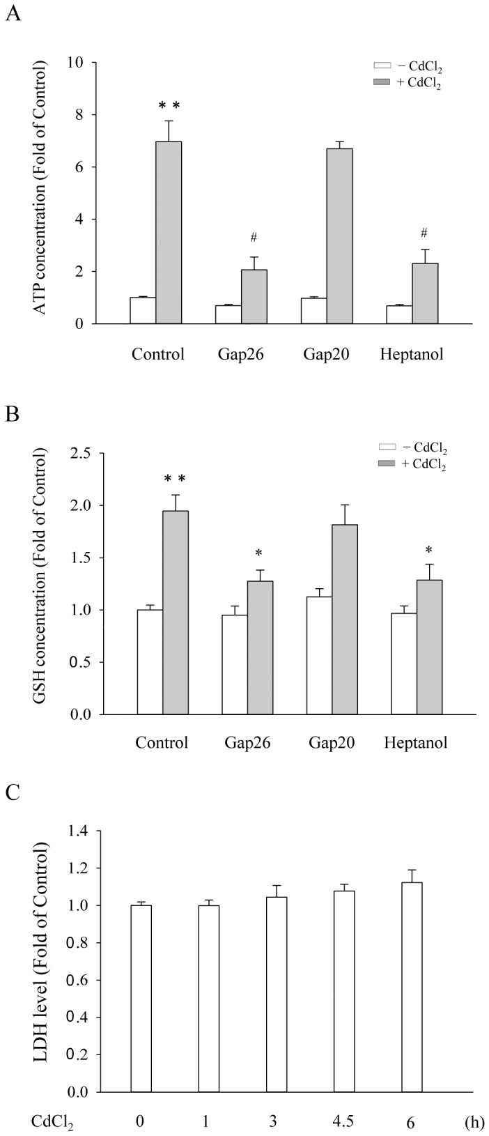
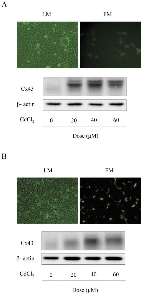
Principal findings: Incubation of Cx43-transfected LLC-PK1 cells with Cd(2+) led to an increased expression of Cx43. This effect of Cd(2+) was tightly associated with JNK activation. Inhibition of JNK abolished the elevation of Cx43. Further analysis revealed that the changes of JNK and Cx43 were controlled by GSH. Supplement of a membrane-permeable GSH analogue GSH ethyl ester or GSH precursor N-acetyl-cystein abrogated the effects of Cd(2+) on JNK activation and Cx43 expression. Indeed, Cd(2+) induced extracellular release of GSH. Blockade of Cx43 hemichannels with heptanol or Cx43 mimetic peptide Gap26 to prevent the efflux of GSH significantly attenuated the Cx43-elevating effects of Cd(2+).

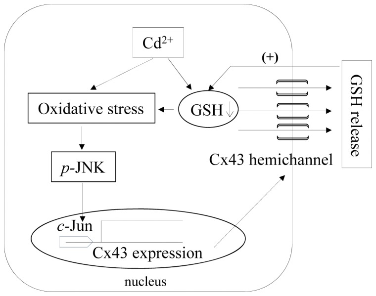
Conclusions: Collectively, our results thus indicate that Cd(2+)-induced upregulation of Cx43 is through activation of nonjunctional Cx43 hemichannels. Our findings thus support the existence of a hemichannel-mediated self-regulation of Cx43 and provide novel insights into the molecular mechanisms of Cx43 expression and function.


Conflict of interest statement

Competing Interests: The authors have declared that no competing interests exist.

Figures

Figure 1
Figure 1. Effects of Cd2+ on Cx43 expression in Cx43-LLC-PK1 cells.
(A–C) Time- and dose-dependent effects of Cd2+ on Cx43 protein levels. Cx43-LLC-PK1 cells were treated with 35 μM CdCl2 for the indicated duration (A) or exposed to the indicated concentrations of CdCl2 for 6 h (C). The cellular protein was extracted and subjected to Western blot analysis of Cx43. In Western blot (A), Cx43 was detected at the molecular near 70 kDa (upper band) and 43 kDa, representing EGFP-tagged and untagged Cx43, respectively. P0, P1 and P2 denoted the nonphophorylated, phosphorylated and hyper-phosphorylated Cx43. (B) Densitometric analysis of Cx43 expression shown in A. Results were expressed as induction relative to the basal level of Cx43 (mean ± S.D., n = 3). # p<0.01 versus untreated control. (D) Effect of Cd2+ on Cx43 mRNA expression. Cx43-LLC-PK1 cells were exposed to 35 μM CdCl2 for the indicated duration. Cellular RNA was extracted and subjected to Northen blot analysis of Cx43. The level of GAPDH was shown as a loading control. (E) Effect of Cd2+ on Cx43-EGFP distribution. LLC-PK1 cells permanently transfected with a vector encoding Cx43-EGFP were exposed to 35 μM CdCl2. The expression and localization of Cx43-EGFP at different time points following Cd2+ stimulation were shown. Note the obviously increased expression of Cx43-EGFP and shift of fusion protein from cell membrane (arrow head) to perinuclear region (white arrow) after incubation with Cd2+.
Figure 2
Figure 2. Involvement of JNK activation in the induction of Cx43 protein by Cd2+.
(A–B) Cd2+-induced p-JNK and c-Jun activation. LLC-PK1 cells were exposed to 35 μM Cd2+ for the indicated time (A) or various concentrations of Cd2+ (B) for 6 h. The cellular protein was extracted and subjected to Western blot analysis of phosphorylated (p) JNK, c-Jun and Cx43. The level of β-actin was taken as loading control. (C–F) Suppression of Cd2+-elicited increase of p-JNK, c-Jun and Cx43 levels by a JNK inhibitor. LLC-PK1 cells were pretreated with 50 μM SP600125 for 30 min before exposing them to 35 μM Cd2+ for 6 h. Cellular proteins were analyzed for p-JNK, c-Jun, Cx43 and β-actin (C). (D–F) Densitometric analysis of p-JNK, c-Jun and Cx43 expression shown in C. Results were expressed as relative induction compared with the basal level (mean ± S.D., n = 3). * p<0.05 versus Cd2+ alone, and ** <0.01 versus untreated control.
Figure 3
Figure 3. Effects of GSH-upregulating agents on Cd2+-elicited Cx43 expression.
Cx43-LLC-PK1 cells were pretreated with 2 mM GSHee or 2 mM NAC for 1 h and exposed to 35 μM Cd2+ for additional 6 h. Cellular protein was subjected to Western blot analysis for p-JNK, c-Jun, Cx43 and β-actin (A). Densitometric results (B–D) were expressed as induction relative to the basal level of p-JNK, c-Jun and Cx43, respectively (mean ± S.D., n = 3). # p<0.01 versus Cd2+ alone, and ** <0.01 versus untreated control.
Figure 4
Figure 4. Influence of gap junctions (GJs) blocker on Cx43 expression and JNK activation.
(A) Cx43-LLC-PK1 cells were pretreated with GJs blocker, 4 mM heptanol for 30 min before exposing them to 35 μM Cd2+ for 6 h. Cellular proteins were analyzed for p-JNK, c-Jun, Cx43 and β-actin. (B–D) Densitometric analysis of p-JNK, c-Jun and Cx43 expression shown in A. Results were expressed as relative induction compared with the basal level (mean ± S.D., n = 3). * p<0.05 versus Cd2+ alone, and ** <0.01 versus untreated control.
Figure 5
Figure 5. Effects of various connexin (Cx) mimetic peptides on Cx43 and p-JNK, c-Jun levels.
(A) Cx43-LLC-PK1 cells were pretreated with various Cx mimetic peptides, Gap20, and Gap26 at the concentration of 100 μM for 1 h, and then exposed to 35 μM Cd2+ in the presence of the peptides for additional 6 h. Cellular protein was subjected to Western blot analysis for p-JNK, c-Jun, Cx43 and β-actin. Densitometric results (B–D) were expressed as induction relative to the basal level of p-JNK, c-Jun and Cx43, respectively (mean ± S.D., n = 3). # p<0.01 and * p<0.05 versus Cd2+ alone, and ** <0.01 versus untreated control.
Figure 6
Figure 6. Blockade of Cd2+-induced ATP and GSH release by GJs inhibitor and Cx mimetic peptides.
(A–B) Cx43-LLC-PK1 cells were pretreated with 4 mM heptanol for 30 min or with Cx43 mimetic peptides Gap20 and Gap26 at the concentration of 100 μM for 1 h, and then exposed to 35 μM Cd2+ in the presence of the pretreated agents for additional 6 h ATP and GSH concentration in culture medium was measured. The data were expressed as the fold induction against basal level (mean ± S.E., n = 4). # p<0.01 and * p<0.05 versus Cd2+ alone, and ** <0.01 versus untreated control. (C) Time course effects of Cd2+ on LDH release. Cx43-LLC-PK1 cells were treated with 35 μM CdCl2 for the indicated duration. The supernatants were collected and assayed for LDH activity. The data were expressed as fold of zero point control (mean ± S.E., n = 4).
Figure 7
Figure 7. Effects of Cd2+ on Cx43 expression in the mixed culture containing both Cx43-positive and Cx43-null LLC-PK1 cells.
(A) Cx43-LLC-PK1 cells and LLC-PK1 cells were cultured in 1∶4 proportions. The number and localization of Cx43-EGFP positive cells under fluorescent microscope in the mixed culture is shown (upper panel, images under light and fluorescent microscope, x200). The mixed culture was exposed to the indicated concentrations of CdCl2 for 6 h. The cellular protein was extracted and subjected to Western blot analysis of Cx43 (lower panel). (B) LLC-PK1 cells were transiently transfected with a wild-type Cx43 gene or EGFP1 vector for 36 h. The number and distribution of EGFP-positive and negative cells under fluorescent microscope are shown and used for estimation of transfection efficiency by comparing with total cells under light microscope and fluorescent microscope (upper panel). The cells transiently transfected with a wild-type Cx43 gene were exposed to the indicated concentrations of CdCl2 for 6 h. Cellular protein was subjected to Western blot analysis for Cx43 (lower panel).
Figure 8
Figure 8. Schematic diagram illustrating Cx43 hemichannel-mediated self-regulation of Cx43 under the stimulation of Cd2+.
Induction of oxidative stress and depletion of GSH by Cd2+ results in the opening of Cx43-hemichannels, leading to the efflux of the major antioxidant GSH. The loss of GSH exaggerates oxidative stress, promoting JNK activation and JNK-mediated Cx43 mRNA expression. The increased synthesis of Cx43 might enhance the formation of nonjunctional hemichannels, further worsening the loss of GSH. The vicious autoregulation loop between GSH and hemichannels provides a molecular mechanism for the hemichannel-mediated regulation of Cx43.

References

    1. Saez JC, Berthoud VM, Branes MC, Martinez AD, Beyer EC (2003) Plasma membrane channels formed by connexins: their regulation and functions. Physiol Rev 83: 1359–1400. - PubMed
    1. Yao J, Zhu Y, Morioka T, Oite T, Kitamura M (2007) Pathophysiological roles of gap junction in glomerular mesangial cells. J Membr Biol 217: 123–130. - PubMed
    1. Yao J, Oite T, Kitamura M (2009) Gap junctional intercellular communication in the juxtaglomerular apparatus. Am J Physiol Renal Physiol 296: F939–946. - PubMed
    1. Berthoud VM, Beyer EC (2009) Oxidative stress, lens gap junctions, and cataracts. Antioxid Redox Signal 11: 339–353. - PMC - PubMed
    1. Upham BL, Trosko JE (2009) Oxidative-dependent integration of signal transduction with intercellular gap junctional communication in the control of gene expression. Antioxid Redox Signal 11: 297–307. - PMC - PubMed

Publication types

MeSH terms