. 2013 Jul 15;22(14):2047-56.
doi: 10.1089/scd.2012.0615. Epub 2013 Apr 6.

Allogeneic mesenchymal stem cell therapy for bisphosphonate-related jaw osteonecrosis in Swine

Affiliations

Allogeneic mesenchymal stem cell therapy for bisphosphonate-related jaw osteonecrosis in Swine

Yunsheng Li et al. Stem Cells Dev. .

Abstract

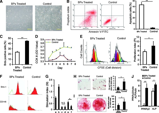
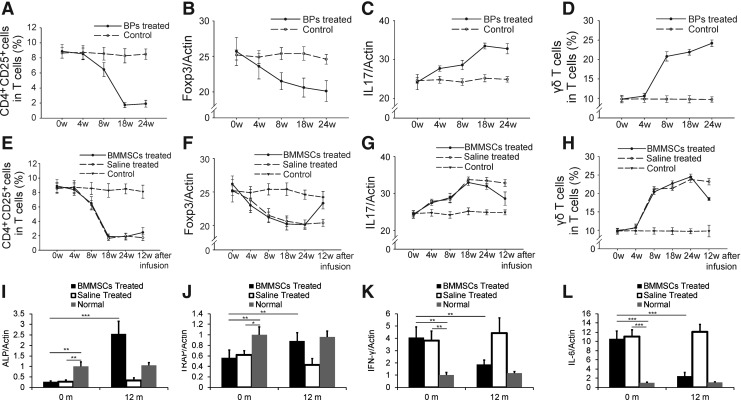
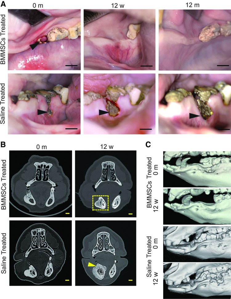
Bisphosphonates (BPs), which are used to treat a variety of clinical disorders, have the side effect of jawbone necrosis. Currently, there is no reliable treatment for BP-related osteonecrosis of the jaw (BRONJ) due to a lack of understanding of its pathogenesis. To investigate the pathogenesis of BRONJ and observe the treatment effect of bone marrow mesenchymal stem cell (BMMSC) transplantation, we established a preclinical animal model of BRONJ in miniature pigs (minipigs). After treatment with zoledronic acid, the clinical and radiographic manifestations of BRONJ could be observed in minipigs after first premolar extraction. The biological and immunological properties of BMMSCs were impaired in the BP-treated minipigs. Moreover, the ratio of Foxp3-positive regulatory T-cells (Tregs) in peripheral blood decreased, and interleukin (IL)-17 increased in the serum of BP-treated minipigs. After allogeneic BMMSC transplantation via intravenous infusion, mucosal healing and bone reconstruction were observed; IL-17 levels were reduced; and Tregs were elevated. In summary, we established a clinically relevant BRONJ model in minipigs and tested a promising allogeneic BMMSC-based therapy, which may have potential clinical applications for treating BRONJ.


Figures

FIG. 1.
FIG. 1.
Generation of a minipig model of bisphosphonate (BP)-related osteonecrosis of the jaw (BRONJ). (A) Schematic illustration of the timeline of the procedures conducted in this study. Eight minipigs were intravenously injected with 4 mg zoledronic acid (ZA) alone once every 2 weeks. Three minipigs were injected with physiological saline as controls. Clinical assessments and computed tomography (CT) examinations were performed for all animals before BP delivery, 24 weeks before tooth extraction, after jaw bone necrosis, and 12 weeks after bone marrow mesenchymal stem cell (BMMSC) infusion. Jaw bone tissue samples were taken for histopathological analysis from all BRONJ minipigs and untreated normal controls before allogeneic BMMSC infusion and 12 weeks after the infusion. (B) Four weeks after extraction, the clinical examination showed incomplete mucosal healing (black arrowhead) in all pigs in the BP treatment group, but no pigs in the untreated group (scale bar=1 cm). (C) Eight weeks after extraction, necrotic bone was exposed in the oral cavity of BP-treated minipigs (black arrowhead), but not in the untreated group (scale bar=1 cm). (D) After tooth extraction, destruction and enlargement of the cortical bone were seen in the BRONJ model on the CT scan. Both osteolysis and osteosclerosis were observed in the necrotic jaw bone (yellow arrowhead) (scale bar=1 cm). (E) The CT scan showed a healed alveolar socket (yellow frame) in the control group (scale bar=1 cm). (F) The 3D images of CT scan showed destruction of the bone in the BP-treated group. (G) Scanning electronic microscopy (SEM) analysis showed collapsed collagen fibers in the necrotic jaw bone of the BP-treated group and normal collagen of the control group at baseline. Transmission electron microscopy (TEM) revealed pyknosis of the nuclei (upper panel) and empty bone lacuna (lower panel). (H) Hematoxylin and eosin (H&E) staining of the minipig BRONJ model showed the presence of fibrosis and inflammatory infiltrate areas and necrotic bone (NB) areas with empty lacunae and fibrosis (scale bar=50 μm). (I) Histological analysis of normal swine jawbones showed regular arrangement of the lamina and active osteocytes in lacunae (scale bar=50 μm). (J) The area of fibrosis and inflammatory infiltrate over the tissue area were significantly higher in the jawbones of minipigs with BRONJ (***P<0.001). (K, L) Trichrome staining of BRONJ (K) and normal (L) swine jawbones showing necrotic bone and a lack of new bone (osteoid, red staining) in the jaws from minipigs with BRONJ (scale bar=200 μm). (M) The area of osteoid over the tissue area was significantly decreased in the jawbones of minipigs with BRONJ (**P<0.01).
FIG. 2.
FIG. 2.
Impairment of biological and immunological functions of BMMSCs in the BP-treated minipigs. (A) BMMSCs in the BP group could hardly adhere compared with controls 1 week after isolation (scale bar=2.0 mm). (B) Annexin V staining for apoptosis showed that ∼43% of BMMSCs from the BRONJ group were positive for the early or late stages of apoptosis, but fewer apoptotic cells (4.14%±0.6%) were found in the untreated control group (**P<0.01). (C) Quantified BrdU-positive cells were significantly decreased in the BP-treated groups (**P<0.01). (D) CCK-8 tests showed a decreased proliferative ability of cells in the BP groups compared with the untreated group. (E) The carboxyfluorescein succinimidyl ester (CFSE) test indicated a declined proliferation index (*P<0.05) in the treatment group compared with the untreated control group. (F) There was no significant difference between the treatment and untreated control groups for both the Stro-1 and CD-146 markers (Stro-1: 64.38%±1.19% vs. 64.46%±0.93%; CD-146: 58.89%±0.42% vs. 55.17%±0.81%, both P>0.05). (G) In mixed lymphocyte reaction, when tested with CCK-8, there was no difference in the stimulation index between the BP and control groups (0.19%±3.2% vs. 0.21%±4.3%, P>0.05), when mixed PBLs and BMMSCs, in the presence of phytohemagglutinin (PHA), the stimulation index increased in the BP groups (0.57%±9.6% vs. 0.22%±6.5%, *P<0.05) (a: PBLs; b: PBLs+PHA; c: PBLs+BMMSCs of BP groups; d: PBLs+BMMSCs of control group; e: PBLs+PHA+BMMSCs of BP groups; f: PBLs+PHA+BMMSCs of control group). (H) The differentiation potential of BMMSCs stained with Oil red O or (I) alizarin red S (scale bar=50 μm) is shown; BMMSCs from BP-treated animals have significantly lower adipogenic differentiation and osteogenic differentiation potential (**P<0.01). (J) Q-PCR showed that PPARγ2 and alkaline phosphatase (ALP) were expressed. Adipose potential, as well as osteogenic potential, was significantly different between the treatment and untreated control groups (**P<0.01; *P<0.05).
FIG. 3.
FIG. 3.
Immunological changes in the BP-treated minipigs and bone remodeling and immunoregulation improvement after BMMSC infusion. (A) CD4+CD25+ T-cells were significantly decreased in the peripheral blood from the BP-treated groups at 24 weeks after the BP treatment compared with the untreated control group (P<0.05). (B) BP treatment also resulted in slightly reduced Foxp3+ levels in the peripheral blood at 24 weeks after BP treatment (P=0.05). (C) interleukin (IL)-17 levels in the peripheral blood in the BP-treated groups were increased compared with the untreated control group at 24 weeks after BP treatment (P<0.05). (D) The levels of γδT cells in the BP groups were significantly increased compared with the control group at 24 weeks after BP treatment (P<0.05). At 12 weeks after BMMSC transplantation, the level of CD4+CD25+ T-cells recovered (P<0.05) (E); the level of Foxp3 mRNA tended to increase in BRONJ minipigs (P<0.05) (F); and IL-17 levels were also reduced after treatment (P<0.05) (G). The level of γδT cells in peripheral blood decreased in the BMMSC group (P<0.05) (H). The expression of ALP (I) and TRAP (J) increased 12 months after BMMSC treatment, whereas the levels of IFN-γ (K) and IL-6 (L) decreased (*P<0.05; **P<0.01; ***P<0.001). IFN, interferon; TRAP, tartrate-resistant acid phosphatase.
FIG. 4.
FIG. 4.
Allogeneic BMMSC transplantation cured BRONJ in the minipig model. (A) The open alveolar socket with NB in the minipig model of BRONJ before BMMSC infusion (black arrowhead) (scale bar=1 cm). BRONJ minipigs that received BMMSC infusions showed healing with complete soft tissue 12 weeks and 12 months after infusion (scale bar=1 cm), whereas the exposure of NB in minipigs with BRONJ before saline infusion was still present with a wider exposure in the same minipigs with BRONJ (black arrowhead) 12 weeks and 12 months after saline infusion (scale bar=1 cm). (B) The CT image showing the necrotic right mandible of minipigs with BRONJ before BMMSC infusion and the disappearance of necrotic bone and healing of the BRONJ 12 weeks after BMMSC infusion (yellow frame) (scale bar=1 cm); more NB was found in the alveolar process (yellow arrowhead) and the lingual side of the right mandible 12 weeks after saline infusion (scale bar=1 cm). (C) Three-dimensional images of the CT scan also showed healing of the bony defects compared with the saline-treated group in the BMMSC-treated group 12 weeks after treatment.
FIG. 5.
FIG. 5.
Histological observation of bone reconstruction in the BMMSC-treated BRONJ minipig model. (A) H&E staining showed new bone formation in the NB area 12 weeks and 12 months after BMMSC infusion (scale bar=300 μm), and NB was still present in minipigs with BRONJ treated with saline (scale bar=300 μm). (B) Positive staining of Y-chromosomes (red) in regenerated bone of the BRONJ region (yellow arrowhead). (C–F) Bone remodeling of the mandible in minipigs after treatment with BMMSCs. Osteoclasts (C, black arrowhead) and osteoblasts (E, green arrowhead) were observed in H&E-stained sections 12 weeks after BMMSC infusions (bar=50 μm). Means of osteoclast/field (D) and osteoblast/field (F) in H&E-stained sections were also significantly higher in the BMMSC-treated group **P<0.01. (G) Trichrome staining revealed new bone formation (osteoid, red staining) in the collagen matrix (scale bar=300 μm) of the group treated with BMMSCs 12 months postinfusion, whereas minipigs with BRONJ treated with saline showed no marked new bone formation (scale bar=300 μm). (H) TRAP staining showed that the osteoclasts (black arrowhead) were observed in the group treated with BMMSCs 12 months postinfusion, whereas minipigs with BRONJ treated with saline showed few osteoclasts (bar=50 μm). A significant increase in the osteoid area (I) and osteoclasts (J) over the tissue area in the group treated with BMMSCs compared with the group treated with saline (***P<0.001).

References

    1. Ruggiero SL. Mehrotra B. Rosenberg TJ. Engroff SL. Osteonecrosis of the jaws associated with the use of bisphosphonates: a review of 63 cases. J Oral Maxillofac Surg. 2004;62:527–534. - PubMed
    1. Burr DB. Allen MR. Mandibular necrosis in beagle dogs treated with bisphosphonates. Orthod Craniofac Res. 2009;12:221–228. - PMC - PubMed
    1. American Association of Oral, Maxillofacial Surgeons. American Association of Oral and Maxillofacial Surgeons position paper on bisphosphonate-related osteonecrosis of the jaws. J Oral Maxillofac Surg. 2007;65:369–376. - PubMed
    1. Ruggiero SL. Dodson TB. Assael LA. Landesberg R. Marx RE. Mehrotra B. American Association of Oral and Maxillofacial Surgeons position paper on bisphosphonate-related osteonecrosis of the jaws. J Oral Maxillofac Surg. 2009;67:2–12. - PubMed
    1. Allen MR. Burr DB. The pathogenesis of bisphosphonate-related osteonecrosis of the jaw: so many hypotheses, so few data. J Oral Maxillofac Surg. 2009;67:61–70. - PubMed

Publication types

MeSH terms

LinkOut - more resources