. 2013 May;280(9):2027-41.
doi: 10.1111/febs.12226. Epub 2013 Apr 8.

Cancer stem-like cell properties are regulated by EGFR/AKT/β-catenin signaling and preferentially inhibited by gefitinib in nasopharyngeal carcinoma

Affiliations

Cancer stem-like cell properties are regulated by EGFR/AKT/β-catenin signaling and preferentially inhibited by gefitinib in nasopharyngeal carcinoma

Lei Ma et al. FEBS J. 2013 May.

Abstract

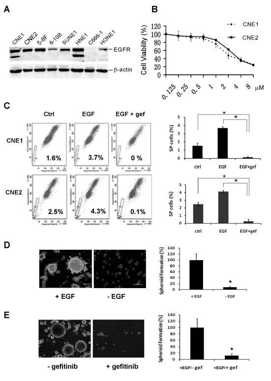
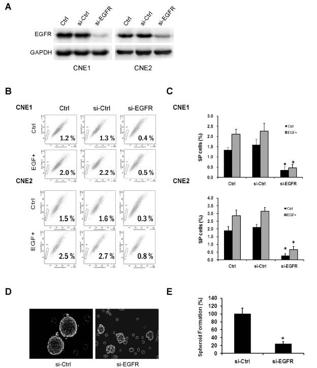
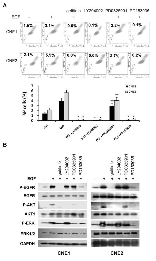
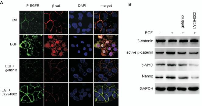
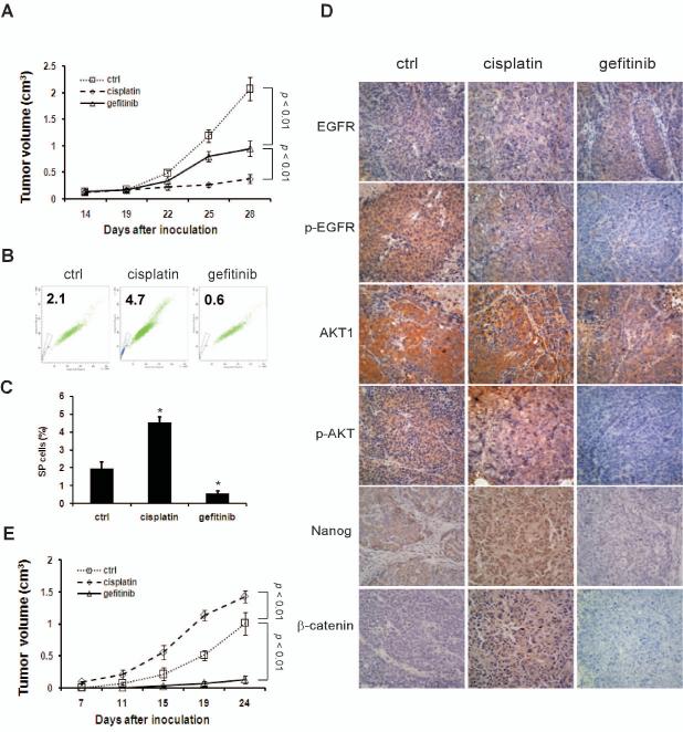
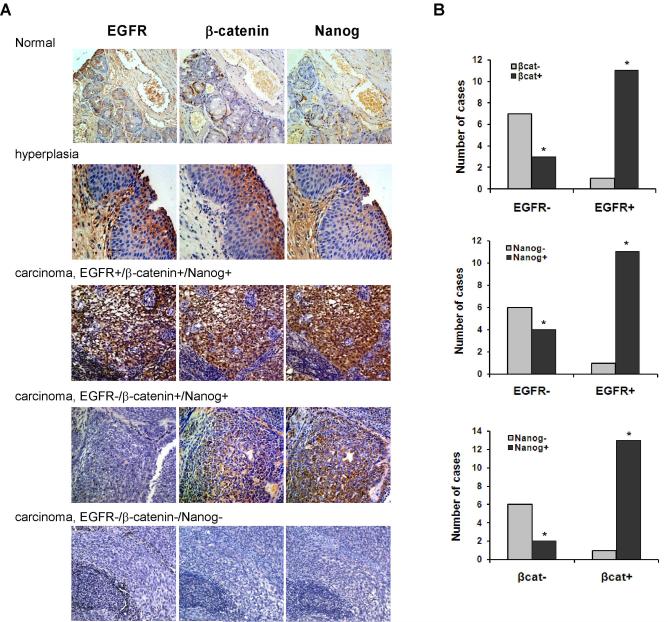
We report that the epidermal growth factor receptor (EGFR) pathway plays a critical role in regulating cancer stem-like cells (CSCs) in nasopharyngeal carcinoma (NPC), one of the most common malignant tumors in Southeast Asia. Effects of EGFR on maintaining CSCs are mainly mediated by AKT signaling, and β-catenin is responsible for governing CSC properties in response to EGFR/AKT activation. Significantly, CSCs are enriched by cisplatin and decreased by gefitinib in NPC xenograft models. Upon reimplantation in secondary mice, tumor cells derived from cisplatin-treated mice grew rapidly, whereas regrowth of tumor cells from gefitinib-treated mice was severely diminished. We further demonstrate that expression of EGFR correlates with expression of β-catenin and Nanog in primary tumor specimens from NPC patients. These findings provide mechanistic and preclinical evidence supporting the use of gefitinib alone or in combination with a chemotherapeutic agent in first-line therapy for patients with NPC. In addition, our results suggest that targeting β-catenin represents a rational clinical modality for patients whose tumors harbor activated EGFR or AKT.


Figures

Fig. 1
Fig. 1
Inhibition of CSCs by gefitinib in NPC. (A) Expression levels of EGFR in 8 NPC cell lines. (B) Antiproliferative effects of gefitinib in CNE1 and CNE2 cell lines. Cells were treated with various concentrations of gefitinib for 72 h, and cell viability was determined by MTT assay. Error bars indicate standard deviations. The experiment was conducted three times with similar results. (C) Inhibition of side population (SP) by gefitinib (1 μM) in NPC cells. Data are presented as mean ± SD (n = 3). *, P< 0.01. (D) EGF dependence of tumor spheroid formation of CNE2 cells. (E) Inhibition of tumor spheroid formation by gefitinib. CNE2 cells derived from primary tumor spheroids were cultured in suspension growth medium containing EGF and bFGF. Gefitinib was added at 2 μM. Bar = mean ± SD (n = 3). *, P< 0.01. Scale bars: 100 μm.
Fig. 2
Fig. 2
Knockdown of EGFR with siRNA diminishes CSC properties. (A) CNE1 and CNE2 cells were transfected with control siRNA or siRNA targeting EGFR followed by Western blot analysis. In both cell lines, exposure to EGFR siRNA substantially reduced EGFR expression. (B and C) After transfected with control siRNA or EGFR siRNA, CNE1 and CNE2 cells were treated with or without EGF (50 ng/mL). Knockdown of EGFR reduced the percentages of the SP of CNE1 and CNE2 at both basal levels and after EGF stimulation. Data are presented as mean ± SD (n = 3). *, P< 0.01. (D and E) Depletion of EGFR suppressed tumor spheroid formation of CNE2 cells. Bars are means ± SD (n = 3). *, P< 0.01.
Fig. 3
Fig. 3
EGFR/PI3K/AKT pathway regulates CSC phenotype in NPC. (A) CNE1 and CNE2 cells were treated with indicated inhibitors for 24 h followed by FACS SP analyses. EGF (50 ng/mL)-induced SP cells were diminished by EGFR inhibitor (either 1 μM of gefitinib or 0.5 μM PD153035) or PI3K inhibitor LY294002 (15 μM), but not by Mek inhibitor PD0325901 (10 μM). Data are presented as mean ± SD (n = 3). *, P< 0.01; **, P> 0.05. (B) Cells were treated as described in (A) followed by Western blot analysis. EGF-induced phosphorylation of EGFR, AKT and ERK1/2 was blocked by gefitinib or PD153035.
Fig. 4
Fig. 4
The effect of EGFR on the CSC phenotype is mediated through β-catenin signaling. (A) In untreated CNE2 cells, β-catenin was located in cytoplasmic membranes and cytoplasm. Treatment with EGF (50 ng/mL) resulted in increased β-catenin nuclear staining, which was reversed by either gefitinib (1 μM) or LY294002 (15 μM). (B) Western blot analysis showed that elevated expression of active β-catenin, c-myc, and Nanog in response to EGF (50 ng/ml) can be reversed by either gefitinib (1 μM) or LY294002 (15 μM).
Fig. 5
Fig. 5
Knockdown of β-catenin inhibits CSC properties. (A) CNE1 and CNE2 cells were infected with control shRNA lentivirus or β-catenin shRNA. In both cell lines, β-catenin protein was markedly reduced following expression of β-catenin shRNA. (B) Infection with β-catenin shRNA lentivirus reduced the percentage of SP cells at the basal level and after EGF (50 ng/mL) stimulation. Bar = mean ± SD (n = 3). *, P< 0.01. (C) Compared with cells infected with the control shRNA (sh-GFP) lentivirus, cells infected with sh-β-catenin lentivirus exhibited reduced tumor spheroid formationin the absence or presence of gefitinib. Bar = mean ± SD (n = 3). *, P< 0.01. Scale bars: 100 μm.
Fig. 6
Fig. 6
Inhibition of CSCs by gefitinib in NPC xenografts. (A) Nude mice harboring CNE2 cell xenografts were treated with saline, gefitinib, or cisplatin for 2 weeks as described in Materials and methods. Graphs represent mean volume ± SD (n = 6). P< 0.01. (B and C) Cells dissociated from primary xenografts were subjected to FACS analyses. Percentages of SP cells were significantly decreased by gefitinib and increased by cisplatin. Bar = mean ± SD (n = 3). *, P< 0.01. (D) Immunohistochemical staining of primary tumors from the mouse model. Photographs were taken at ×200 magnification. (E) Cells from primary xenografts were reimplanted into nude mice for development of secondary tumors. Gefitinib abrogated tumor regeneration in secondary mice. Graphs represent mean volume ± SD (n = 6).
Fig. 7
Fig. 7
Correlation of expression of EGFR, β-catenin, and Nanog in human NPC samples. (A) Immunohistochemical studies of representative NPC specimens showing staining of EGFR, β-catenin, and Nanog. Normal and hyperplastic nasopharyngeal tissues were included as controls. Photographs were taken at ×200 magnification. (B) Expression of EGFR is associated with that of both β-catenin (P< 0.01) and Nanog (P< 0.05), and immunoreactivity of β-catenin is associated with that of Nanog (P< 0.01).

References

    1. Normanno N, De Luca A, Bianco C, Strizzi L, Mancino M, Maiello MR, Carotenuto A, De Feo G, Caponigro F, Salomon DS. Epidermal growth factor receptor (EGFR) signaling in cancer. Gene (Amsterdam) 2006;366:2–16. - PubMed
    1. Mangelberger D, Kern D, Loipetzberger A, Eberl M, Aberger F. Cooperative Hedgehog-EGFR signaling. Front Biosci. 2012;17:90–9. - PMC - PubMed
    1. Eccles SA. The epidermal growth factor receptor/Erb-B/HER family in normal and malignant breast biology. Int J Dev Biol. 2011;55:685–96. - PubMed
    1. Reynolds BA, Weiss S. Generation of Neurons and Astrocytes from Isolated Cells of the Adult Mammalian Central-Nervous-System. Science. 1992;255:1707–1710. - PubMed
    1. Reynolds BA, Tetzlaff W, Weiss S. A Multipotent Egf-Responsive Striatal Embryonic Progenitor-Cell Produces Neurons and Astrocytes. Journal of Neuroscience. 1992;12:4565–4574. - PMC - PubMed

Publication types

MeSH terms