. 2013 Apr;140(7):1475-85.
doi: 10.1242/dev.087601. Epub 2013 Mar 5.

Coronary veins determine the pattern of sympathetic innervation in the developing heart

Affiliations

Coronary veins determine the pattern of sympathetic innervation in the developing heart

Joseph Nam et al. Development. 2013 Apr.

Abstract

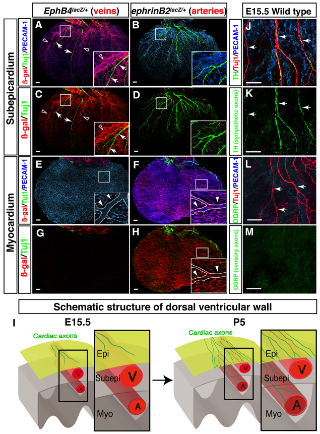
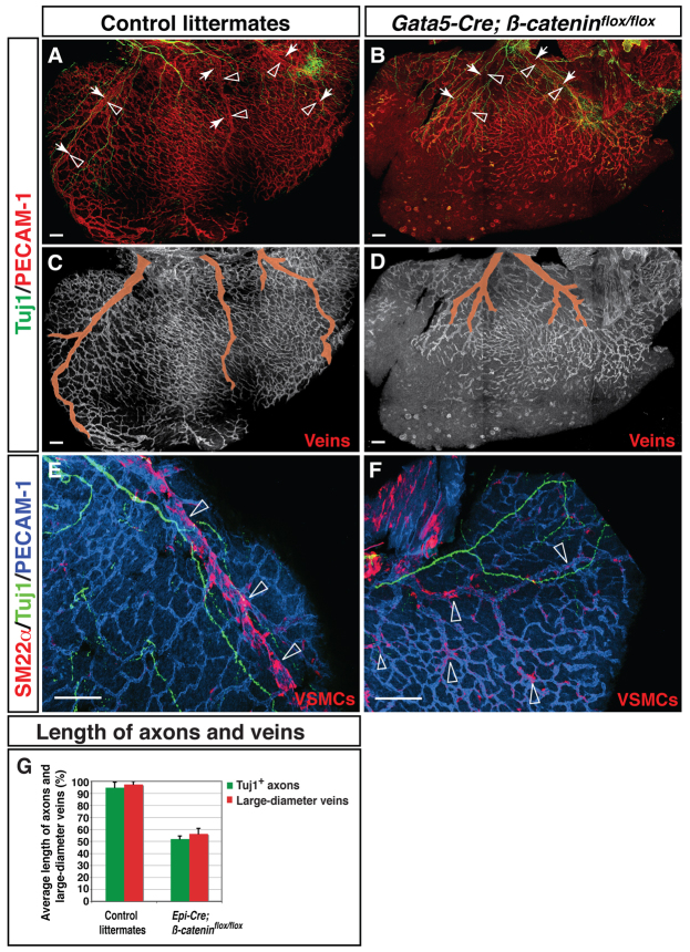
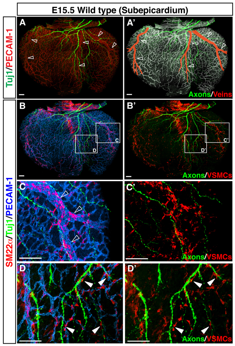
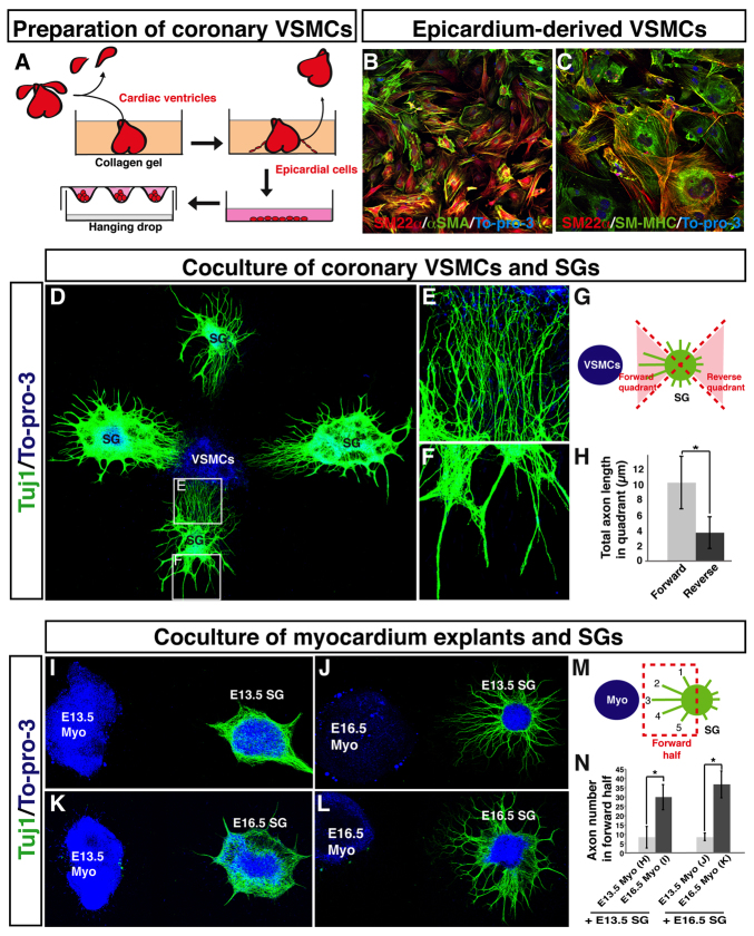
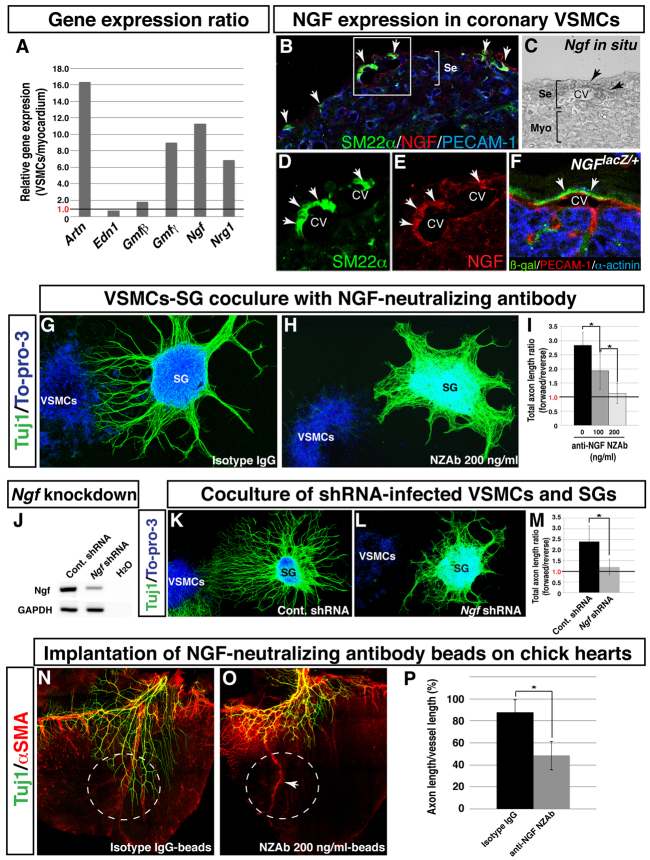
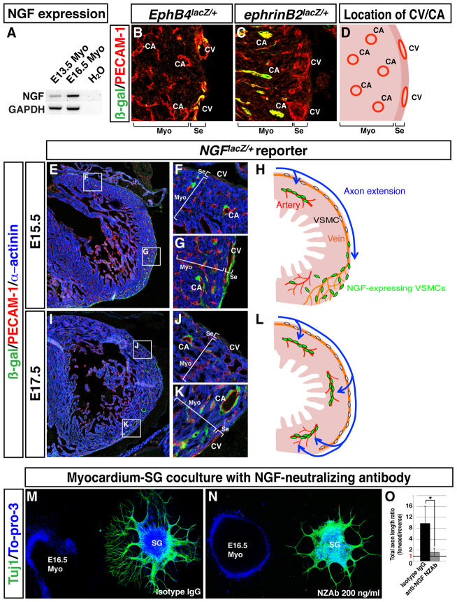
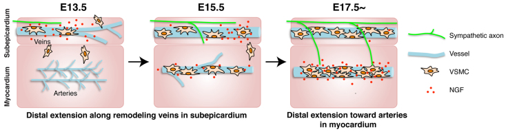
Anatomical congruence of peripheral nerves and blood vessels is well recognized in a variety of tissues. Their physical proximity and similar branching patterns suggest that the development of these networks might be a coordinated process. Here we show that large diameter coronary veins serve as an intermediate template for distal sympathetic axon extension in the subepicardial layer of the dorsal ventricular wall of the developing mouse heart. Vascular smooth muscle cells (VSMCs) associate with large diameter veins during angiogenesis. In vivo and in vitro experiments demonstrate that these cells mediate extension of sympathetic axons via nerve growth factor (NGF). This association enables topological targeting of axons to final targets such as large diameter coronary arteries in the deeper myocardial layer. As axons extend along veins, arterial VSMCs begin to secrete NGF, which allows axons to reach target cells. We propose a sequential mechanism in which initial axon extension in the subepicardium is governed by transient NGF expression by VSMCs as they are recruited to coronary veins; subsequently, VSMCs in the myocardium begin to express NGF as they are recruited by remodeling arteries, attracting axons toward their final targets. The proposed mechanism underlies a distinct, stereotypical pattern of autonomic innervation that is adapted to the complex tissue structure and physiology of the heart.


Figures

Fig. 1.
Fig. 1.
Cardiac nerves align with large diameter coronary vessels in the dorsal ventricular wall of the developing heart. (A-D′) The dorsal face of the cardiac ventricles of an E15.5 mouse embryo. Whole-mount double immunofluorescence confocal microscopy using antibodies to the pan-endothelial marker PECAM1 (A-D, red; A′-D′, white) and the neuronal marker class III β-tubulin (TUJ1, green) reveals that TUJ1+ cardiac axons (A,A′) follow three remodeled large diameter coronary vessels: the right cardiac vein (RCV), the medial branch of left cardiac vein (mLCV), and the lateral branch of left cardiac vein (lLCV) (A, open arrowheads; A′, pseudocolored in red). Magnified images (B-D′) of the boxed regions in A clearly demonstrate the physical proximity of TUJ1+ cardiac axons (B-D′, arrows) and large diameter vessels (B-D, open arrowheads; B′-D′, pseudocolored in red). (E,F) Quantification of nerve-vessel association. In 17 radial sections of the dorsal ventricular wall (termed A-Q, see A′), axons most commonly project to regions containing RCV, mLCV or lLCV. *P<0.01 (Student’s t-test); n=7; error bars indicate s.e.m. RV, right ventricle; LV, left ventricle. Scale bars: 100 μm.
Fig. 2.
Fig. 2.
Cardiac sympathetic axons associate with large diameter coronary veins within the subepicardial layer of the dorsal ventricular wall. (A-H) The dorsal ventricular walls of E15.5 EphB4taulacZ/+ (A,C,E,G; the venous marker EPHB4) or ephrinB2taulacZ/+ (B,D,F,H; the arterial marker ephrin B2) hearts are shown. Whole-mount triple immunofluorescence confocal microscopy was performed with antibodies to PECAM1 (A,B,E,F, blue), TUJ1 (A-H, green) and β-galactosidase (A-H, red). Boxed regions in A-F,H are magnified in insets. (A-D) The subepicardium. Coronary veins expressing EphB4taulacZ are clearly visible in EphB4taulacZ/+ embryos (A,C). However, arteries expressing ephrinB2taulacZ are barely detectable in ephrinB2taulacZ/+ embryos (B,D). TUJ1+ cardiac axons (A,C, arrows) associate with EPHB4+ large diameter veins (A,C, open arrowheads). (E-H) The myocardium. Coronary arteries expressing ephrinB2taulacZ cover the deeper layer in ephrinB2taulacZ/+ embryos (F,H). One large diameter artery runs from the base towards the apex of the ventricle (E,F,H, insets, arrowheads). EphB4taulacZ-expressing veins are barely detectable in EphB4taulacZ/+ embryos (E,G). TUJ1+ cardiac axons are also not detected in the myocardial layer (E-H). (I) Schematic illustrating sympathetic innervation of the developing heart. By E15.5, coronary veins develop to form large diameter branches within the subepicardial layer (Subepi), where cardiac axons initiate distal axon extension. Coronary arteries develop separately, in the myocardial layer (Myo). By P5, cardiac axons extend into the myocardial layer (see supplementary material Fig. S2). These axons innervate large diameter coronary arteries as final targets. Epi, epicardial layer; V, vein; A, artery. (J-M) Neuronal subtype characterization. E15.5 hearts were labeled with antibodies to the sympathetic neuron marker tyrosine hydroxylase (TH; J,K, green) or the sensory neuron marker calcitonin gene related peptide (CGRP; L,M, green) in addition to PECAM1 (J,L, blue) and TUJ1 (J,L, red). TUJ1+ nerves are mostly TH+, indicating that these axons in the subepicardium are mostly sympathetic nerves (J,K, arrows). CGRP+ sensory innervation is not detectable at E15.5 (L,M, arrows). Scale bars: 100 μm.
Fig. 3.
Fig. 3.
Defective coronary development leads to abnormal sympathetic innervation. (A-D) Dorsal ventricular walls of Gata5-Cre; β-cateninflox/flox mutants (B,D) and control littermates (A,C) at E15.5. Double immunofluorescence confocal microscopy was performed with antibodies to PECAM1 (A,B, red; C,D, white) and TUJ1 (A-D, green). In Gata5-Cre; β-cateninflox/flox mice the pattern of coronary remodeling appears disorganized compared with control littermates (A versus B, open arrowheads; C versus D, pseudocolored in red). The mutants also exhibit abnormal sympathetic innervation (A versus B, arrows). Both large diameter veins and sympathetic axons fail to fully develop in the subepicardium. (E,F) Vascular smooth muscle cell (VSMC) recruitment. Triple staining with antibodies to the VSMC marker SM22α (E,F, red) in addition to PECAM1 (E,F, blue) and TUJ1 (E,F, green) revealed that SM22α+ VSMCs associate less strongly with large diameter veins in these mutants; SM22α+ VSMCs are distributed more uniformly throughout the subepicardium, indicating defects in angiogenic remodeling (E versus F, open arrowheads, G). (G) Quantification of nerve-vessel association. The length of large diameter vessels and of sympathetic axons is significantly affected in Gata5-Cre; β-cateninflox/flox mutants. Length is measured as a percentage of distance from base to apex. Control littermates, n=3; Gata5-Cre; β-cateninflox/flox mutants, n=3; error bars indicate s.e.m. Scale bars: 100 μm.
Fig. 4.
Fig. 4.
VSMCs cover nerve-associated large diameter coronary veins. (A-D′) Visualization of coronary VSMCs in the subepicardium. Whole-mount triple immunofluorescence confocal microscopy was performed with antibodies to the VSMC marker SM22α (B-D′, red) in addition to PECAM1 (A, red; A′, white; B-D, blue) and TUJ1 (green). Magnified images (C-D′) show the boxed regions in B,B′. At E15.5, SM22α+ VSMCs are found predominantly around nerve-associated large diameter coronary veins (A-C′, open arrowheads), and a smaller number of SM22α+ VSMCs are located between remodeled veins (D,D′, arrowheads). Scale bars: 100 μm.
Fig. 5.
Fig. 5.
Fetal epicardium-derived VSMCs promote axon outgrowth from fetal sympathetic ganglia in vitro. (A) Schematic illustrating the preparation of coronary VSMCs. E12.5 or E13.5 cardiac ventricles were cultured in collagen gel for 2 days. The ventricles were removed from the gel, and migrated epicardial cells were harvested and further expanded on a type IV collagen-coated dish. VSMC aggregations were obtained using a hanging drop culture method. (B,C) Primary culture of epicardial-derived VSMCs stained with antibodies to VSMC markers SM22α (B,C, green) and αSMA (B, red), and SM-MHC (C, green), together with the nuclear dye To-pro-3 (blue). (D-H) Co-culture of coronary VSMCs and sympathetic ganglia (SG). E13.5 fetal SG were cultured with VSMC aggregations obtained as in A and stained with anti-TUJ1 antibody (green) and To-pro-3 (blue). Magnified images (E,F) show the boxed regions in D. Note that directional axon outgrowth towards VSMCs was observed (E). The total lengths of axons in the forward and reverse quadrants (G) were calculated using Volocity (H). n=19. (I-N) Co-culture of myocardium explants and SG. SG were cultured with fetal myocardial tissue explants and labeled with anti-TUJ1 antibody (green) and To-pro-3 (blue). E13.5 myocardial tissue explants failed to stimulate axon outgrowth from E13.5 and E16.5 SG explants (I,K), whereas E16.5 myocardial tissue explants successfully induced directional axon outgrowth from both E13.5 and E16.5 SG explants (J,L). The total number of axons in the forward region (M) for each sample was quantified using Volocity (N). E13.5 SG with E13.5 myo, n=7; E13.5 SG with E16.5 myo, n=8; E16.5 SG with E13.5 myo, n=5, E16.5 SG with E16.5 myo, n=6. *P<0.01 (Student’s t-test); error bars indicate s.e.m. Myo, myocardium.
Fig. 6.
Fig. 6.
Coronary VSMCs stimulate directional axon growth by NGF in vitro. (A) Semi-quantitative RT-PCR analysis showing differential expression of neurotrophic factors as indicated between VSMCs and E13.5 myocardial tissues. A ratio exceeding 1.0 indicates that the factor is more highly expressed in VSMCs than in myocardial tissues. (B-F) NGF expression in coronary VSMCs in the subepicardium. Triple immunofluorescence confocal microscopy of E15.5 heart sections was performed using antibodies to SM22α (B,D, green) and NGF (B,E, red) as well as PECAM1 (B, blue). Magnified images (D,E) show the boxed region in B. NGF expression was detected in venous VSMCs in the subepicardium (B,D,E, arrows). In situ hybridization with Ngf RNA probes on E15.5 heart section shows that Ngf mRNA is expressed in coronary veins in the subepicardium (C, arrows). The NGF-expressing cells were also detected in coronary veins by triple staining of E15.5 NGFlacZ/+ reporter heart sections with antibodies for β-gal (green), PECAM1 (red) and the myocardial cell marker α-actinin (blue) (F, arrows). CV, coronary vein; Se, subepicardium; Myo, myocardium. (G-I) VSMC-mediated directional axon outgrowth is attenuated by anti-NGF neutralizing antibody (NZAb). E13.5 SG were cultured with VSMC aggregations in the presence of control isotype IgG (G) or 200 ng/ml anti-NGF NZAb (H), and were labeled with anti-TUJ1 antibody (green) and To-pro-3 (blue). Anti-NGF NZAb selectively inhibited directional axon outgrowth as compared with control isotype IgG. The level of inhibition varies with the concentration of anti-NGF NZAb (I). The total lengths of axons in the forward and reverse quadrants (see Fig. 5M) were calculated using Volocity. The ratios of the total lengths of axons in the forward versus reverse quadrants are shown (I). A ratio above 1.0 indicates directional axon outgrowth towards VSMC aggregates. *P<0.05 (Student’s t-test); isotype IgG, n=8; 100 ng/ml anti-NGF NZAb, n=5; 200 ng/ml anti-NGF NZAb, n=6. (J-M) Ngf-deficient VSMCs fail to induce directional axon outgrowth. VSMCs were infected with a control or Ngf shRNA lentivirus during primary epicardial culture. The expression levels of Ngf were assessed by RT-PCR analysis (J). E13.5 SG were cultured with control VSMCs (K) or Ngf-deficient VSMCs (L) and labeled with anti-TUJ1 antibody (green) and To-pro-3 (blue). Ngf-deficient VSMCs failed to induce preferential directional axon outgrowth. Directional outgrowth was quantified as in Fig. 5G (M). *P<0.05 (Student’s t-test); control shRNA-infected VSMCs, n=3; Ngf shRNA-infected VSMCs, n=5. (N-P) Effect of anti-NGF NZAb on coronary VSMC-mediated sympathetic axon growth in chick embryonic hearts. Control isotype IgG-soaked beads or anti-NGF NZAb-soaked beads were implanted on the dorsal surface of E6 chick hearts. After 4 days of incubation, the hearts were dissected for whole-mount double immunofluorescence confocal microscopy using antibodies to TUJ1 (N,O, green) and αSMA (N,O, red). Control beads have no effect (N, white dashed circle) and anti-NGF NZAb beads show no distal extension of sympathetic axons (O, white dashed circle). Distal axon extension was quantified (P). *P<0.01 (Student’s t-test); isotype IgG, n=5; anti-NGF NZAb, n=5. Note that anti-NGF NZAb beads do not inhibit the formation of large diameter coronary vessels (O, arrow). Error bars indicate s.e.m.
Fig. 7.
Fig. 7.
Dynamic expression of NGF in VSMCs from the subepicardium to the myocardium is responsible for myocardial innervation. (A) RT-PCR analysis of E13.5 and E16.5 myocardium explants. (B-D) Location of EPHB4+ coronary veins (B) and ephrin B2+ arteries (C) in the cardiac ventricles. Sections of EphB4lacZ/+ (B) and ephrinB2lacZ/+ (C) hearts were stained with antibodies for β-gal (green) and PECAM1 (red). The relative location of coronary veins (CV) and arteries (CA) in heart ventricle is shown in D. (E-L) NGF expression analysis on NGFlacZ/+ reporter heart sections. E15.5 (E-G) or E17.5 (I-K) heart sections of NGFlacZ/+ reporter embryos were stained with antibodies for β-gal (green), PECAM1 (red) and α-actinin (blue). Magnified images (F,G,J,K) show the boxed regions in E,I. Schematic models illustrating dynamic expression of NGF in VSMCs in parallel with sympathetic axon extension at E15.5 (H) and E17.5 (L). (M-O) SG and myocardium co-culture with anti-NGF NZAb. E16.5 SG were cultured with E16.5 myocardial explants in the presence of 200 ng/ml isotype IgG (M) or 200 ng/ml anti-NGF NZAb (N). The culture was stained with anti-TUJ1 antibody (green) and To-pro-3 (blue). (O) Directional axon growth was quantified as in Fig. 5G. *P<0.02 (Student’s t-test); isotype IgG, n=5; 200 ng/ml anti-NGF NZAb, n=5; error bars indicate s.e.m. CV, coronary vein; CA, coronary artery; Myo, myocardium; Se, subepicardium.
Fig. 8.
Fig. 8.
A two-step process is responsible for sympathetic innervation of the developing heart. Model for sympathetic axon innervation of the developing mouse heart. At E13.5, coronary veins undergo angiogenic remodeling in the subepicardium. Venous VSMCs transiently secrete NGF as they associate with remodeled veins. Sympathetic axons start to extend into the subepicardium. By E15.5, NGF expression in venous VSMCs proceeds distally in parallel with vascular remodeling, and axons extend along newly formed veins. Some arterial VSMCs begin to express NGF in the myocardium. By E17.5, which is the stage when sympathetic innervation penetrates the myocardium, NGF expression is detected only in arterial VSMCs.

References

    1. Cai C. L., Martin J. C., Sun Y., Cui L., Wang L., Ouyang K., Yang L., Bu L., Liang X., Zhang X., et al. (2008). A myocardial lineage derives from Tbx18 epicardial cells. Nature 454, 104–108 - PMC - PubMed
    1. Carmeliet P., Tessier-Lavigne M. (2005). Common mechanisms of nerve and blood vessel wiring. Nature 436, 193–200 - PubMed
    1. Ciszek B., Skubiszewska D., Ratajska A. (2007). The anatomy of the cardiac veins in mice. J. Anat. 211, 53–63 - PMC - PubMed
    1. Enomoto H., Crawford P. A., Gorodinsky A., Heuckeroth R. O., Johnson E. M., Jr, Milbrandt J. (2001). RET signaling is essential for migration, axonal growth and axon guidance of developing sympathetic neurons. Development 128, 3963–3974 - PubMed
    1. Francis N., Farinas I., Brennan C., Rivas-Plata K., Backus C., Reichardt L., Landis S. (1999). NT-3, like NGF, is required for survival of sympathetic neurons, but not their precursors. Dev. Biol. 210, 411–427 - PubMed

Publication types

MeSH terms