. 2013 Apr;140(7):1445-56.
doi: 10.1242/dev.071878. Epub 2013 Mar 5.

SOX2 maintains the quiescent progenitor cell state of postnatal retinal Muller glia

Affiliations

SOX2 maintains the quiescent progenitor cell state of postnatal retinal Muller glia

Natalia Surzenko et al. Development. 2013 Apr.

Abstract

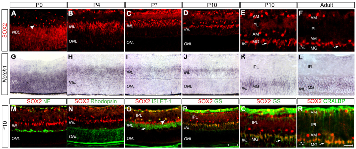
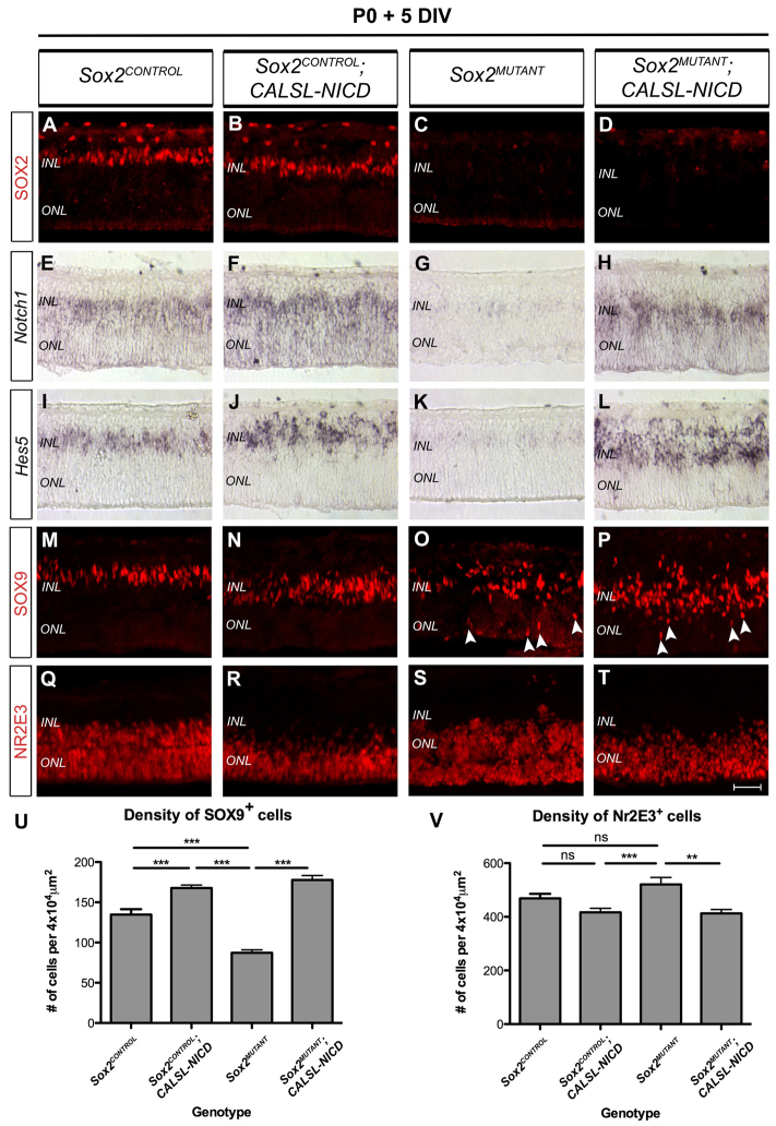
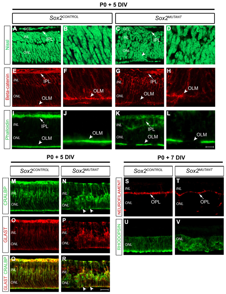
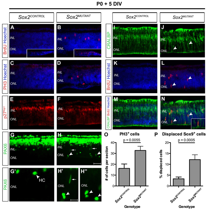
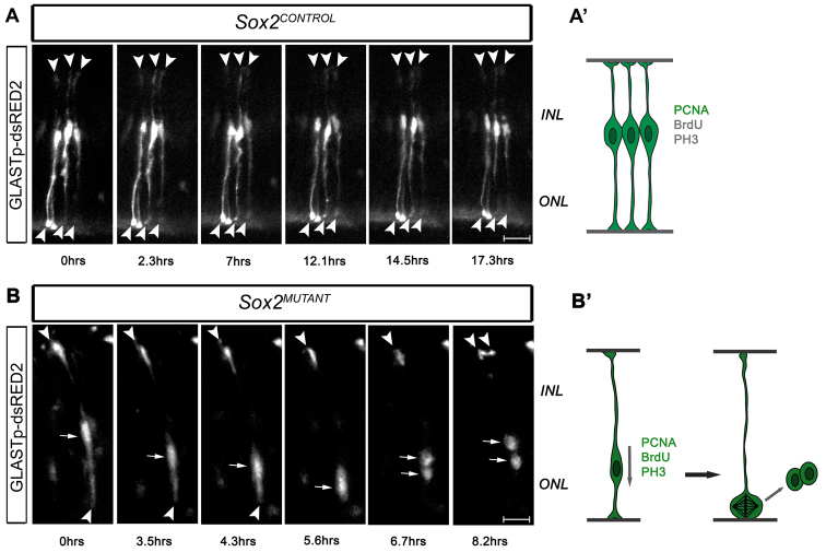
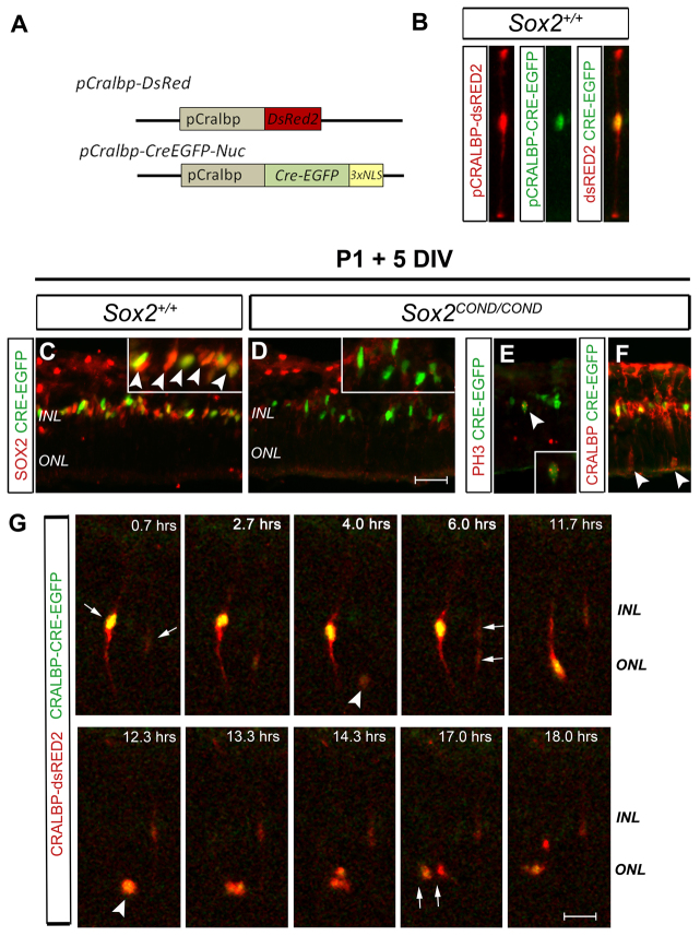
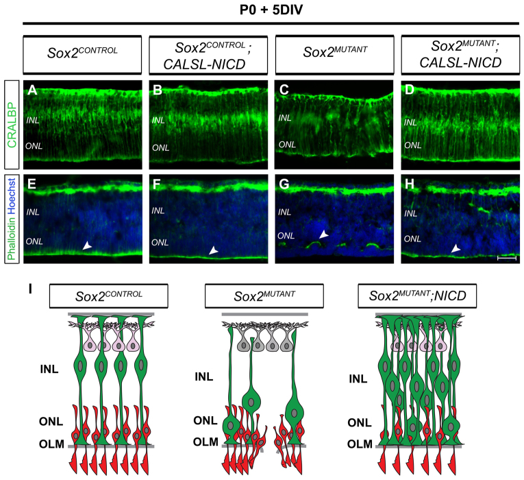
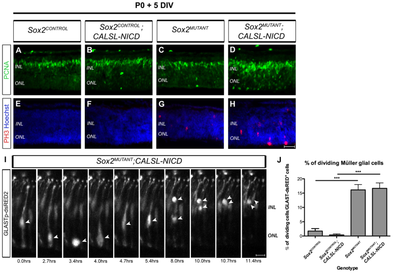
Within discrete regions of the developing mammalian central nervous system, small subsets of glia become specialized to function as neural stem cells. As a result of their self-renewal and neurogenic capacity, these cells later serve to replenish neurons and glia during persistent or injury-induced adult neurogenesis. SOX2, an HMG box transcription factor, plays an essential role in the maintenance of both embryonic and adult neural progenitors. It is unclear, however, which biological mechanisms regulated by SOX2 are required for neural stem cell maintenance. In this study, we address this question through genetic analysis of SOX2 function in differentiating postnatal Müller glia, a cell type that maintains neurogenic capacity in the adult retina. By utilizing molecular analysis and real-time imaging, we show that two progenitor characteristics of nascent Müller glia - their radial morphology and cell cycle quiescence - are disrupted following conditional genetic ablation of Sox2 in the mouse postnatal retina, leading to Müller cell depletion and retinal degeneration. Moreover, we demonstrate that genetic induction of the Notch signaling pathway restores Müller glial cell identity to Sox2 mutant cells, but does not secure their quiescent state. Collectively, these results uncouple the roles of SOX2 and the Notch signaling pathway in the postnatal retina, and uncover a novel role for SOX2 in preventing the depletion of postnatal Müller glia through terminal cell division.


Figures

Fig. 1.
Fig. 1.
SOX2 and NOTCH1 are expressed in postnatal retinal progenitor cells and in Müller glia. (A-F) Expression of SOX2 in the mouse retina was assessed by immunohistochemistry at the indicated ages. Arrowhead (A), amacrine cell; arrows (E,F), Müller glia (MG). (G-L) Notch1 expression was examined by in situ hybridization. (M-R) SOX2 does not colocalize with NF (M) or rhodopsin (N). SOX2 is co-expressed with ISLET1 in amacrine cells (O, arrowhead) but not in bipolar cells (O, arrow). SOX2 colocalizes with GS (P, higher magnification in Q) and CRALBP (R) in MG at P10 (P,Q) and in the adult retina (R). AM, amacrine cells; GS, glutamine synthetase; IPL, inner plexiform layer; INL, inner nuclear layer; NBL, neuroblast layer; NF, neurofilament; ONL, outer nuclear layer. Scale bars: 65 μm in P for A-D,G-J,M-P; 30 μm in R for E,F,K,L,Q,R.
Fig. 2.
Fig. 2.
Loss of NOTCH1 activity and disorganization of MG in Sox2MUTANT retinas. (A-D) SOX2 expression is not detected in Sox2MUTANT (C) and Sox2MUTANT;CALSL-NICD (D) TM-treated retinas cultured for 5 days. (E-L) Expression (in situ hybridization) of Notch1 (E-H) and Hes5 (I-L) is lost in Sox2MUTANT (G,K) retinas and is restored by NICD activity in Sox2MUTANT;CALSL-NICD retinas (H,L). (M-P) SOX9-expressing MG are displaced to the ONL in both Sox2MUTANT (O, arrowheads) and Sox2MUTANT;CALSL-NICD (P, arrowheads) retinas. (Q-T) NR2E3-expressing rod photoreceptor precursors are reduced in number in Sox2CONTROL;CALSL-NICD (R) and Sox2MUTANT;CALSL-NICD (T) retinas, and are disorganized in Sox2MUTANT retinas (S). (U,V) Total densities of SOX9-positive (U) and NR2E3-positive (V) cells were quantified on sections of TM-treated retinas of the indicated genotypes. ***P<0.0001; ns, not significant. Error bars indicate s.e.m. Sox2CONTROL refers to Sox2+/+;CAGGCre-ER™; Sox2MUTANT refers to Sox2COND/COND;CAGGCre-ER™. TM, 4-hydroxytamoxifen. Scale bar: 45 μm in A-T.
Fig. 3.
Fig. 3.
Defects in retinal lamination and MG morphology in Sox2MUTANT retinas. (A-L) Immunohistochemical analysis of sectioned retinas reveals disorganization of lamination in Sox2MUTANT retinas treated with TM and cultured for 5 days. B,D,F,H,J,L are high magnification images of A,C,E,G,I,K. (A-D) In Sox2MUTANT retinas, the ONL, INL and GCL are disorganized and cells are displaced beyond the OLM (C, arrowhead) and IPL (C, arrow), as shown by Nissl staining (A,B versus C,D). (E-H) Whereas β-catenin is enriched in adherens junctions of the IPL (E, arrow) and OLM (E,F, arrowheads) of Sox2CONTROL retinas, β-catenin staining in the IPL (G, arrow) and OLM (G,H, arrowheads) of Sox2MUTANT retinas is discontinuous. (I-L) Breaks in the IPL (arrows) and OLM (arrowheads) of Sox2MUTANT retinas are visualized by the uneven distribution of F-actin (phalloidin), which is normally enriched in the IPL (I,K, arrows) and OLM (I-L, arrowheads). (M-R) Immunostaining against CRALBP (M,N) and GLAST (O,P) reveals the radial morphology of MG in Sox2CONTROL retinas (M,O,Q), whereas MG in Sox2MUTANT retinas are disorganized and displaced to the ONL (N,P,R, arrowheads). (S,T) The OPL, marked by NF, is disorganized in Sox2MUTANT (T) compared with Sox2CONTROL (S) retinas cultured for 7 days. (U,V) Rod photoreceptors form rosette structures in Sox2MUTANT (V) compared with Sox2CONTROL (U) retinas cultured for 7 days. GCL, ganglion cell layer; OLM, outer limiting membrane. Scale bars: 40 μm in R for M-R; 15 μm in L for B,D,F,H,J,L.
Fig. 4.
Fig. 4.
MG in Sox2MUTANT retinas express mitotic cell cycle markers. (A-D) The number of cells incorporating BrdU (A,B) and exhibiting PH3 (C,D) in the central and peripheral regions of Sox2MUTANT retinas is increased (insets in A,B). (E,F) Expression of p27Kip1 in the INL of Sox2MUTANT retinas (F) is decreased compared with Sox2CONTROL retinas. (G-H″) PAX6 is not detected in the ONL, but marks amacrine cells, MG and horizontal cells (G′, arrowhead) in the INL (G,G′) in Sox2CONTROL retinas. In Sox2MUTANT retinas, PAX6 is found in elongated cells (H,H′, arrows) and in cells with a round morphology in the ONL (H,H′,H″, arrowheads). (I-N) CRALBP-positive MG do not incorporate BrdU in Sox2CONTROL retinas (I,K,M), but are BrdU positive in Sox2MUTANT retinas (J,L,N, arrowheads, inset). (O) The number of PH3-positive cells is higher in Sox2MUTANT than Sox2CONTROL retinas. (P) The percentage of SOX9-expressing MG displaced to the ONL is increased in Sox2MUTANT (12.16±2.23) compared with Sox2CONTROL (3.31±0.84) retinas (two-tailed t-test; P=0.0005). Data are mean ± s.e.m. HC, horizontal cell. Scale bars: 45 μm in H for A-H,I-N; 25 μm in H′ for G′-H″.
Fig. 5.
Fig. 5.
Cell division of MG in Sox2MUTANT retinas. (A,A′) Time-lapse imaging of GLASTp-dsRED2-expressing cells in slices of TM-treated Sox2CONTROL retinas at day 3-4 of culture illustrates MG radial morphology, maintenance of apical and basal cellular processes (arrowheads) and limited cell body movement (illustrated in A′). (B,B′) A GLASTp-dsRED2-labeled MG imaged in the Sox2MUTANT retina migrates to the ONL and undergoes cell division, followed by separation of the two daughter cells (arrows) and splitting of the basal cellular process (arrowheads) (illustrated in B′). Image series were collected on 200 μm retinal sections every 40 minutes over 8-18 hours. Scale bars: 30 μm in A; 20 μm in B.
Fig. 6.
Fig. 6.
Mosaic ablation of Sox2 leads to ectopic cell division of nascent MG. (A) pCRALBP-dsRED2 and pCRALBP-CreEGFP-Nuc DNA constructs. (B) CRALBP-dsRED2 colocalizes with CRALBP-CreEGFP in MG in retinas co-electroporated at P1 and cultured for 5 days. (C) In Sox2+/+ retinas, CRALBP-CreEGFP is co-expressed with SOX2 in the INL (C, arrowheads). (D) SOX2 is not detected in EGFP-positive cells in Sox2COND/COND retinas. (E,F) A subset of CRALBP-CreEGFP-expressing MG in Sox2COND/COND retinas exhibit PH3 (E, arrowhead, inset) and are displaced to the ONL (F, arrowheads). (G) CRALBP-CreEGFP/CRALBP-dsRED2 double-labeled MG (arrows) in Sox2COND/COND retinas undergo interkinetic nuclear migration and cell division (see supplementary material Movie 5). Arrowheads indicate mitotic MG at the apical retinal boundary. Confocal images (G) were collected from 200 μm retinal sections every 40 minutes over an 18-hour period. Scale bars: 45 μm in C-F; 35 μm in G.
Fig. 7.
Fig. 7.
Activation of Notch signaling in Sox2MUTANT;CALSL-NICD retinas restores MG cell morphology and retinal architecture. (A-D) CRALBP reveals restoration of MG morphology and retinal architecture in Sox2MUTANT;CALSL-NICD retinas, as compared with Sox2MUTANT retinas (C versus D). (E-H) The integrity of the OLM is restored in Sox2MUTANT;CALSL-NICD retinas (H, arrowhead) compared with Sox2MUTANT retinas (G, arrowhead). (I) Comparison of MG morphology and retinal organization between Sox2CONTROL, Sox2MUTANT and Sox2MUTANT;CALSL-NICD retinas. MG in Sox2MUTANT retinas are reduced in number and are displaced to the ONL. NICD activity restores the number of MG, but not their INL position. MG, green; rod photoreceptors, red; amacrine cells, gray, pink. Scale bar: 45 μm in A-H.
Fig. 8.
Fig. 8.
NICD activity does not restore the quiescent state of MG in Sox2MUTANT;CALSL-NICD retinas. (A-D) PCNA-expressing cells are present in both the INL and ONL of Sox2MUTANT;CALSL-NICD retinas (D). (E-H) PH3-positive cells are found in Sox2MUTANT (G) and Sox2MUTANT;CALSL-NICD (H) retinas. (I) Daughter cell resulting from a dividing GLASTp-dsRED2-labeled MG (arrowheads) re-establishes radial morphology in Sox2MUTANT;CALSL-NICD retina. The image series were collected on 200 μm sections every 40 minutes over a 12-hour period. (J) The percentage of dividing GLASTp-dsRED2-positive MG is significantly increased in Sox2MUTANT and Sox2MUTANT;CALSL-NICD retinas (P<0.0001) compared with Sox2CONTROL and Sox2CONTROL;CALSL-NICD retinas. ***P<0.0001. Error bars indicate s.e.m. Scale bars: 45 μm in A-H; 30 μm in I.

References

    1. Austin C. P., Feldman D. E., Ida J. A., Jr, Cepko C. L. (1995). Vertebrate retinal ganglion cells are selected from competent progenitors by the action of Notch. Development 121, 3637–3650 - PubMed
    1. Bao Z. Z., Cepko C. L. (1997). The expression and function of Notch pathway genes in the developing rat eye. J. Neurosci. 17, 1425–1434 - PMC - PubMed
    1. Bringmann A., Pannicke T., Grosche J., Francke M., Wiedemann P., Skatchkov S. N., Osborne N. N., Reichenbach A. (2006). Müller cells in the healthy and diseased retina. Prog. Retin. Eye Res. 25, 397–424 - PubMed
    1. Bylund M., Andersson E., Novitch B. G., Muhr J. (2003). Vertebrate neurogenesis is counteracted by Sox1-3 activity. Nat. Neurosci. 6, 1162–1168 - PubMed
    1. Chenn A., Walsh C. A. (2002). Regulation of cerebral cortical size by control of cell cycle exit in neural precursors. Science 297, 365–369 - PubMed

Publication types

MeSH terms

Substances