Review
. 2013 Mar;7(1):11-6.
doi: 10.1007/s12105-013-0428-1. Epub 2013 Mar 5.

The importance of diagnosing NUT midline carcinoma

Affiliations
Review

The importance of diagnosing NUT midline carcinoma

Christopher A French. Head Neck Pathol. 2013 Mar.

Abstract

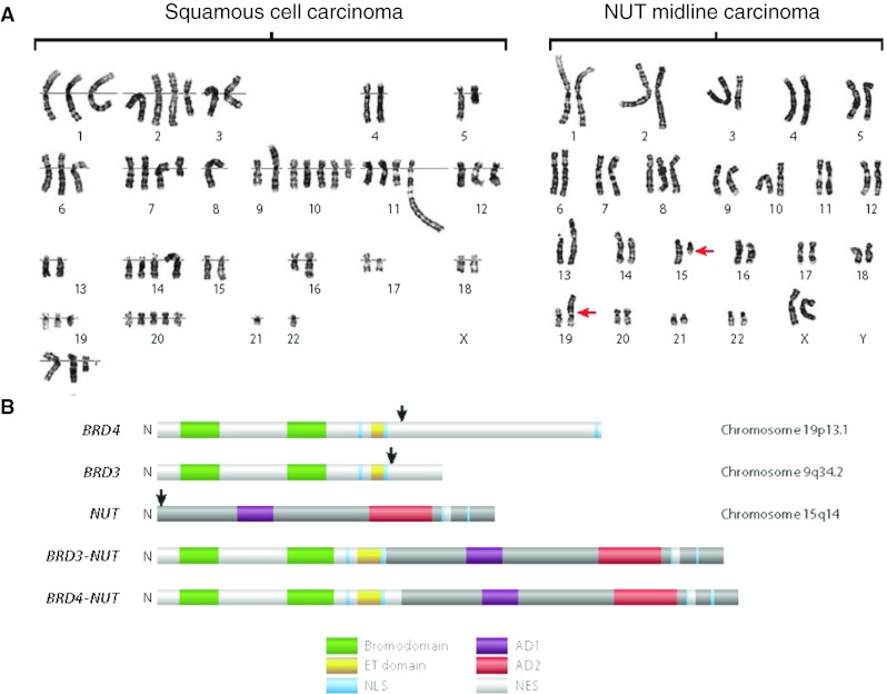
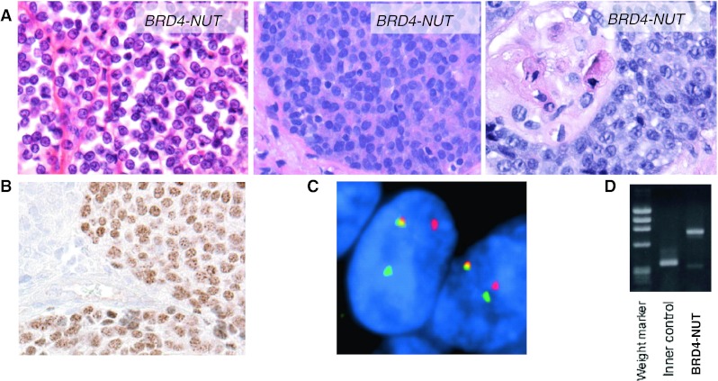
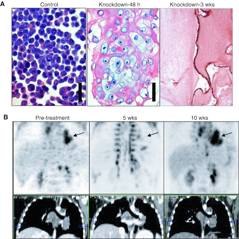
NUT midline carcinoma (NMC) is an aggressive subset of squamous cell carcinoma, genetically defined by rearrangement of the NUT gene. The rearrangements most often take the form of BRD4-NUT fusions, and in a minority of cases, BRD3-NUT or NUT-variant fusions. The simple karyotypes of NMCs, in contrast to the complex ones of typical squamous cell carcinoma, suggest an alternate, genetic shortcut to squamous cancer. Although originally thought to be a disease of the mediastinum, NMC frequently (35 %) arises in the head and neck. Diagnosis is made simply by demonstration of nuclear immunoreactivity to NUT protein, and ancillary studies to characterize the fusion oncogene, though not required for diagnosis, are recommended. The prognosis is dismal, with a 6.7 month median survival, and treatment with conventional chemotherapeutic regimens is ineffective. The oncogenic mechanism of the dual bromodomains and the p300-binding portion of BRD4-NUT is to sequester p300 to localized regions of chromatin, leading to global transcriptional repression and blockade of differentiation. Two therapies which target this mechanism have emerged, including bromodomain inhibitors (BETi) and histone deacetylase inhibitors (HDACi), both of which induce differentiation and growth arrest of NMC cells, both in vitro and in vivo. BETi is available to adults with NMC through a phase I clinical trial, and clinical response to HDACi has been demonstrated in pediatric patients. The emergence of these promising targeted therapies gives hope that NMC may one day be effectively treated and provides a strong rationale for diagnostic testing for NMC.


Figures

Fig. 1
Fig. 1
Genetic aberrations in NUT midline carcinoma (NMC). a Karyotypes of typical non–NUT midline carcinoma (NMC) squamous cell carcinoma (left), compared with (right) NMC. The red arrows denote chromosomal translocation between chromosomes 15 and 19. b Domain structures encoded by BRD-NUT fusions and native component genes. The black arrows indicate the locations of translocation-associated break points. AD1 acidic domain 1, AD2 acidic domain 2, ET extraterminal, NES nuclear export signal, NLS nuclear localization signal. a and b were taken from Annual Reviews of Pathology [7]
Fig. 2
Fig. 2
Diagnosis of NUT midline carcinoma (NMC). a H&E photomicrographs (400×) of three different NMCs reveal monotony and occasional abrupt squamous differentiation. Adapted from Annual Reviews of Pathology [7]. b Immunohistochemistry using a NUT-specific antibody (Cell Signaling Technologies) reveals speckled nuclear staining. c Fluorescent in situ hybridization (FISH) using a dual-color breakapart probe flanking the NUT gene. The split-apart red and green signals indicate rearrangement of the NUT locus. Taken from Cancer Research [2]. d Reverse-transcriptase polymerase chain reaction (RT-PCR) using primers that flank the coding sequence of the BRD4-NUT breakpoint and appropriate controls. Courtesy of Yukichi Tanaka, Mio Tanaka, Toru Horisawa, and Yutaka Saikawa, Kanazawa Medical University, Ishikawa, Japan
Fig. 3
Fig. 3
Targeting NUT midline carcinoma (NMC). a Small interfering RNA (siRNA) knockdown of BRD4-NUT in patient-derived NUT midline carcinoma cells (TC-797) using an siRNA directed against NUT [5] (scale bar 50 μm). Control consists of TC-797 cells 72 h after transfection with scrambled siRNA. Taken from Annual Reviews of Pathology [7]. b P18PF-fluorodeoxyglucose–positron emission tomography and computed tomography scan of the patient’s mediastinal tumor (arrow) reveals arrested growth five weeks following HDACi (vorinostat) treatment, then recurrence five weeks after the patient became intolerant to the drug and ceased taking it. Taken from Cancer Research [16]

References

    1. French CA, Kutok JL, Faquin WC, et al. Midline carcinoma of children and young adults with NUT rearrangement. J Clin Oncol. 2004;22:4135–4139. doi: 10.1200/JCO.2004.02.107. - DOI - PubMed
    1. Bauer D, Mitchell C, Strait K, et al. Clinicopathologic features and long-term outcomes of NUT midline carcinoma. Clin Cancer Res. 2012;18:5773–5779. doi: 10.1158/1078-0432.CCR-12-1153. - DOI - PMC - PubMed
    1. Haack H, Johnson LA, Fry CJ, et al. Diagnosis of NUT midline carcinoma using a NUT-specific monoclonal antibody. Am J Surg Pathol. 2009;33:984–991. doi: 10.1097/PAS.0b013e318198d666. - DOI - PMC - PubMed
    1. French CA, Miyoshi I, Kubonishi I, et al. BRD4-NUT fusion oncogene: a novel mechanism in aggressive carcinoma. Cancer Res. 2003;63:304–307. - PubMed
    1. French CA, Ramirez CL, Kolmakova J, et al. BRD-NUT oncoproteins: a family of closely related nuclear proteins that block epithelial differentiation and maintain the growth of carcinoma cells. Oncogene. 2008;27:2237–2242. doi: 10.1038/sj.onc.1210852. - DOI - PubMed

Publication types

MeSH terms