

Physiological roles of dietary glutamate signaling via gut-brain axis due to efficient digestion and absorption
- PMID: 23463402
- PMCID: PMC3698427
- DOI: 10.1007/s00535-013-0778-1
Physiological roles of dietary glutamate signaling via gut-brain axis due to efficient digestion and absorption
Abstract
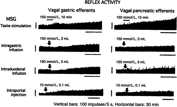
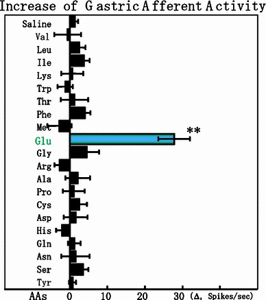
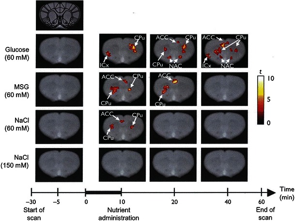
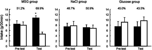
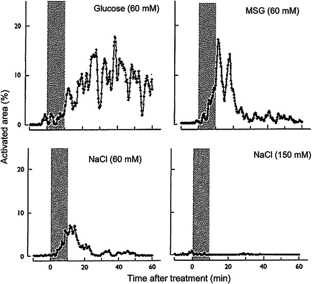
Dietary glutamate (Glu) stimulates to evoke the umami taste, one of the five basic tastes, enhancing food palatability. But it is also the main gut energy source for the absorption and metabolism for each nutrient, thus, only a trace amount of Glu reaches the general circulation. Recently, we demonstrated a unique gut sensing system for free Glu (glutamate signaling). Glu is the only nutrient among amino acids, sugars and electrolytes that activates rat gastric vagal afferents from the luminal side specifically via metabotropic Glu receptors type 1 on mucosal cells releasing mucin and nitrite mono-oxide (NO), then NO stimulates serotonin (5HT) release at the enterochromaffin cell. Finally released 5HT stimulates 5HT3 receptor at the nerve end of the vagal afferent fiber. Functional magnetic resonance imaging (f-MRI, 4.7 T) analysis revealed that luminal sensing with 1 % (w/v) monosodium L-glutamate (MSG) in rat stomach activates both the medial preoptic area (body temperature controller) and the dorsomedial hypothalamus (basic metabolic regulator), resulting in diet-induced thermogenesis during mealing without changes of appetite for food. Interestingly, rats were forced to eat a high fat and high sugar diet with free access to 1 % (w/w) MSG and water in a choice paradigm and showed the strong preference for the MSG solution and subsequently, they displayed lower fat deposition, weight gain and blood leptin. On the other hand, these brain functional changes by the f-MRI signal after 60 mM MSG intubation into the stomach was abolished in the case of total vagotomized rats, suggesting that luminal glutamate signaling contributes to control digestion and thermogenesis without obesity.
Figures
References
-
- Giacometti T. Free and bound glutamate in natural products. In: Filer LJ Jr, Garattini MR, Kare MR, Reynolds WA, Wurtman RJ, editors. Glutamic acid: advances in biochemistry and physiology. New York: Raven Press; 1979. pp. 25–34.
-
- Young VR, Ajami AM. Glutamate: an amino acid of particular distribution. J Nutr. 2000;130(Suppl 4S):892S–900S. - PubMed
-
- Hector MP. Reflexes of salivary secretion. In: Garrett JR, Ekström J, Anderson LC, editors. Neural mechanisms of salivary gland secretion. Basel: Karger; 1999. pp. 196–217.
Publication types
MeSH terms
Substances
LinkOut - more resources
Full Text Sources
Other Literature Sources
