. 2013 Apr 26;288(17):11649-61.
doi: 10.1074/jbc.M112.445304. Epub 2013 Mar 5.

The intracellular interactome of tetraspanin-enriched microdomains reveals their function as sorting machineries toward exosomes

Affiliations

The intracellular interactome of tetraspanin-enriched microdomains reveals their function as sorting machineries toward exosomes

Daniel Perez-Hernandez et al. J Biol Chem. .

Abstract

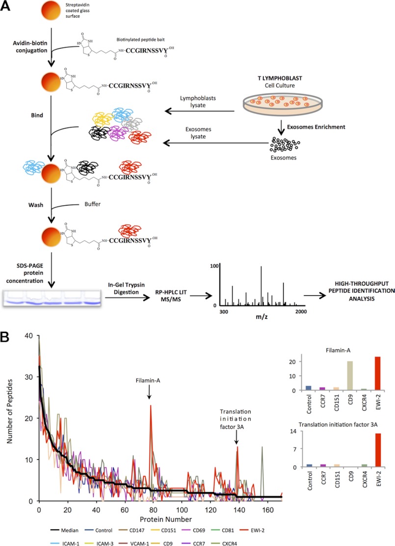
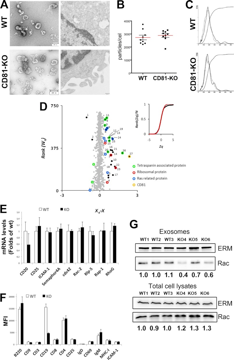
Extracellular vesicles are emerging as a potent mechanism of intercellular communication because they can systemically exchange genetic and protein material between cells. Tetraspanin molecules are commonly used as protein markers of extracellular vesicles, although their role in the unexplored mechanisms of cargo selection into exosomes has not been addressed. For that purpose, we have characterized the intracellular tetraspanin-enriched microdomain (TEM) interactome by high throughput mass spectrometry, in both human lymphoblasts and their derived exosomes, revealing a clear pattern of interaction networks. Proteins interacting with TEM receptors cytoplasmic regions presented a considerable degree of overlap, although some highly specific CD81 tetraspanin ligands, such as Rac GTPase, were detected. Quantitative proteomics showed that TEM ligands account for a great proportion of the exosome proteome and that a selective repertoire of CD81-associated molecules, including Rac, is not correctly routed to exosomes in cells from CD81-deficient animals. Our data provide evidence that insertion into TEM may be necessary for protein inclusion into the exosome structure.


Figures

FIGURE 1.
FIGURE 1.
Scheme of the protocol followed for the high throughput analysis of the interactome of the tetraspanin-enriched microdomains. A, biotinylated, synthetic peptides containing the intracellular regions (C-terminal in all cases except for CD69) of tetraspanins CD9, CD81(depicted in the figure), and CD151, their associated receptors (EWI-2, ICAM-1, and VCAM-1), or different control receptors (CD147, CD69, ICAM-3, CCR7, and CXCR4) were bound to streptavidin-Sepharose beads and incubated with protein extracts from whole cell lysates or exosomes from human primary lymphoblasts. Beads were washed and directly subjected to concentrating SDS-PAGE. Protein bands were trypsin-digested, and the resulting peptides were identified by liquid chromatography in tandem with linear ion trap mass spectrometry. B, peptide counting analysis reveals specific protein interactions with peptide baits. The left plot represents the distribution of the number of peptides identified per each protein in a representative pulldown assay. On the right, the number of peptides from the proteins is indicated by the arrows, which show specific interactions with one or more baits are shown.
FIGURE 2.
FIGURE 2.
Specificity of interactions between TEM receptors and intracellular proteins in cell lysates and exosomes of human primary lymphoblasts. Charts show proteomic analysis (high throughput mass spectrometry) of pulldown assays in whole cell lysates (left) or exosome extracts (right) of primary human lymphoblasts. Data correspond to interaction with α-actinin (A), filamin (B), nucleolin (C), Rac (D), elongation factor 1-α (E), actin (F), and ERMs (G). Western blotting of equivalent pulldowns in whole cell lysates and exosomes confirms these interactions.
FIGURE 3.
FIGURE 3.
Protein interaction networks for ligands of CD81, EWI-2, and ICAM-1. A, intracellular ligands of CD81, EWI-2, or ICAM-1 were manually clustered into groups according to their function using the IPA analysis program. Symbol size is proportional to the number of peptides identified for each protein in the pulldown assays. B, Venn diagram showing the degree of overlap between the sets of proteins interacting with CD81, EWI-2, and ICAM-1 baits.
FIGURE 4.
FIGURE 4.
Protein interaction networks for CD81 and EWI-2 ligands identified in human primary lymphoblast-derived exosomes. A, intracellular ligands of CD81 and EWI-2 were manually clustered into groups according to their function using the IPA analysis program. Symbol size is proportional to the number of peptides identified for each protein in the pulldown assays. B, proportions of proteins in exosomes that were identified as ligands of EWI-2 and CD81 in exosomes. The proportions were calculated on the basis of the number of peptides identified per protein in total exosome lysates.
FIGURE 5.
FIGURE 5.
Lack of CD81 alters the protein composition of exosomes. A, exosomes (left panels) were enriched from lymphoblast culture media as described under “Experimental Procedures” and negatively stained. Cells (right panels) were pelleted and fixed with 2% glutaraldehyde and 4% paraformaldehyde and embedded into epoxy resin. Ultrathin sections were viewed in a Jeol JEM-1010 electron microscope after counterstaining with uranyl acetate and lead citrate. A multivesicular body is shown. B, exosome counts in the supernatants of 7-day cultures of mouse blasts from WT or KO animals by NTA. Data represent the mean ± S.E. of individual measurements. C, NTA profile of exosome samples derived from the supernatants of 7-day cultures of mouse blasts from WT or CD81 KO animals. Two different profiles are overlaid for each sample. D, quantitative proteomics comparison of the compositions of pooled exosome extracts from three WT or three CD81 knock-out mouse lymphoblast cultures by stable isotope labeling with 16O (wild type) or 18O (CD81 KO). The plot shows the distribution of log2 ratios of individual proteins, corrected for the grand mean, and ranked by their statistical weight, which is the inverse of the variance and measures the accuracy of the quantification. Black dots indicate proteins with statistically significant abundance changes with respect to the bulk of nonchanging proteins (gray dots). The quantified proteins are listed in supplemental Data File 4. The numbers indicate the proteins as listed in Table 1. The inset plot shows the cumulative frequency distribution of the standardized variable Zq (black line), which expresses the quantitative data in terms of units of variance, showing the agreement with the expected null hypothesis distribution (red line) and the deviation due to proteins changing their abundance. Quantified proteins from the same ontological categories show a similar pattern of abundance changes. Colored points indicate ribosomal proteins, Ras-related proteins, and membrane proteins associated with tetraspanins with a Zq ≥ |2.4|. E, quantitative RT-PCR analyses of mRNA of selected molecules in 7-day cultures of mouse blasts from three WT animals or three CD81 KO mice (mean ± S.D.) F, flow cytometry analyses of selected receptors in 7-day cultures of mouse blasts from three WT animals or three CD81 KO mice. Data represent the mean fluorescent intensity (MFI) (mean ± S.D.) of the positive cells for each marker. G, Western blot analysis of Rac content in exosomes and total cell lysates from independent lymphoblast cultures from wild-type (WT1–3) or CD81 knock-out (KO4–6) mice. Numbers represent densitometric values of Rac signal corrected with ERM loading and normalized to WT1 sample.

References

    1. Yáñez-Mó M., Barreiro O., Gordon-Alonso M., Sala-Valdés M., Sánchez-Madrid F. (2009) Tetraspanin-enriched microdomains: a functional unit in cell plasma membranes. Trends Cell Biol. 19, 434–446 - PubMed
    1. Charrin S., le Naour F., Silvie O., Milhiet P. E., Boucheix C., Rubinstein E. (2009) Lateral organization of membrane proteins: tetraspanins spin their web. Biochem. J. 420, 133–154 - PubMed
    1. Yáñez-Mó M., Gutiérrez-López M. D., Cabañas C. (2011) Functional interplay between tetraspanins and proteases. Cell. Mol. Life Sci. 68, 3323–3335 - PMC - PubMed
    1. Berditchevski F., Odintsova E. (2007) Tetraspanins as regulators of protein trafficking. Traffic 8, 89–96 - PubMed
    1. Mittelbrunn M., Sánchez-Madrid F. (2012) Intercellular communication: diverse structures for exchange of genetic information. Nat. Rev. Mol. Cell Biol. 13, 328–335 - PMC - PubMed

Publication types