

Recurrent NCOA2 gene rearrangements in congenital/infantile spindle cell rhabdomyosarcoma
- PMID: 23463663
- PMCID: PMC3734530
- DOI: 10.1002/gcc.22050
Recurrent NCOA2 gene rearrangements in congenital/infantile spindle cell rhabdomyosarcoma
Abstract
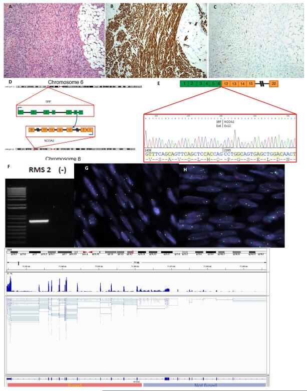
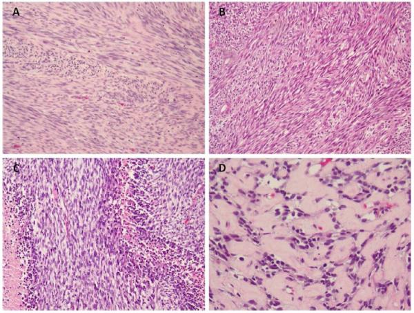
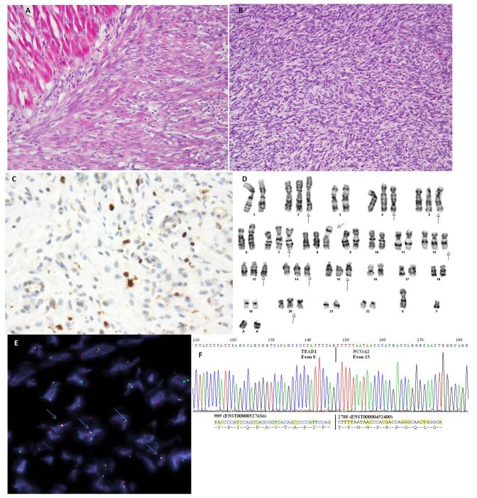
Spindle cell rhabdomyosarcoma (RMS) is a rare form of RMS with different clinical characteristics between children and adult patients. Its genetic hallmark remains unknown and it remains debatable if there is pathogenetic relationship between the spindle cell and the so-called sclerosing RMS. We studied two pediatric and one adult spindle cell RMS by next generation RNA sequencing and FusionSeq data analysis to detect novel fusions. An SRF-NCOA2 fusion was detected in a spindle cell RMS from the posterior neck in a 7-month-old child. The fusion matched the tumor karyotype and was confirmed by FISH and RT-PCR, which showed fusion of SRF exon 6 to NCOA2 exon 12. Additional 14 spindle cell (from 8 children and 6 adults) and 4 sclerosing (from 2 children and 2 adults) RMS were tested by FISH for the presence of abnormalities in NCOA2, SRF, as well as for PAX3 and NCOA1. NCOA2 rearrangements were found in two additional spindle cell RMS from a 3-month-old and a 4-week-old child. In the latter tumor, TEAD1 was identified by rapid amplification of cDNA ends (RACE) to be the NCOA2 gene fusion partner. None of the adult tumors were positive for NCOA2 rearrangement. Despite similar histomorphology in adults and young children, these results suggest that spindle cell RMS is a heterogeneous disease genetically as well as clinically. Our findings also support a relationship between NCOA2-rearranged spindle cell RMS occurring in young childhood and the so-called congenital RMS, which often displays rearrangements at 8q13 locus (NCOA2).
Copyright © 2013 Wiley Periodicals, Inc.
Figures
References
-
- Azakie A, Lamont L, Fineman JR, He Y. Divergent transcriptional enhancer factor-1 regulates the cardiac troponin T promoter. Am J Physiol Cell Physiol. 2005;289:C1522–1534. - PubMed
-
- Calabrese G, Guanciali Franchi P, Stuppia L, Rossi C, Bianchi C, Antonucci A, Palka G. Translocation (8;11)(q12-13;q21) in embryonal rhabdomyosarcoma. Cancer Genet Cytogenet. 1992;58:210–211. - PubMed
-
- Cavazzana AO, Schmidt D, Ninfo V, Harms D, Tollot M, Carli M, Treuner J, Betto R, Salviati G. Spindle cell rhabdomyosarcoma. A prognostically favorable variant of rhabdomyosarcoma. Am J Surg Pathol. 1992;16:229–235. - PubMed
-
- Chen D, Ma H, Hong H, Koh SS, Huang SM, Schurter BT, Aswad DW, Stallcup MR. Regulation of transcription by a protein methyltransferase. Science. 1999;284:2174–2177. - PubMed
-
- Chiles MC, Parham DM, Qualman SJ, Teot LA, Bridge JA, Ullrich F, Barr FG, Meyer WH. Sclerosing rhabdomyosarcomas in children and adolescents: a clinicopathologic review of 13 cases from the Intergroup Rhabdomyosarcoma Study Group and Children’s Oncology Group. Pediatr Dev Pathol. 2004;7:583–594. - PubMed
Publication types
MeSH terms
Substances
Grants and funding
LinkOut - more resources
Full Text Sources
Other Literature Sources
Miscellaneous
