. 2014 Jan;73(1):263-9.
doi: 10.1136/annrheumdis-2012-202726. Epub 2013 Mar 5.

Heightened immune response to autocitrullinated Porphyromonas gingivalis peptidylarginine deiminase: a potential mechanism for breaching immunologic tolerance in rheumatoid arthritis

Affiliations
Free PMC article

Heightened immune response to autocitrullinated Porphyromonas gingivalis peptidylarginine deiminase: a potential mechanism for breaching immunologic tolerance in rheumatoid arthritis

Anne-Marie Quirke et al. Ann Rheum Dis. 2014 Jan.
Free PMC article

Abstract

Background: Rheumatoid arthritis (RA) is characterised by autoimmunity to citrullinated proteins, and there is increasing epidemiologic evidence linking Porphyromonas gingivalis to RA. P gingivalis is apparently unique among periodontal pathogens in possessing a citrullinating enzyme, peptidylarginine deiminase (PPAD) with the potential to generate antigens driving the autoimmune response.

Objectives: To examine the immune response to PPAD in patients with RA, individuals with periodontitis (PD) and controls (without arthritis), confirm PPAD autocitrullination and identify the modified arginine residues.

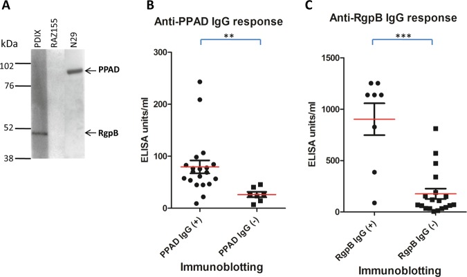
Methods: PPAD and an inactivated mutant (C351A) were cloned and expressed and autocitrullination of both examined by immunoblotting and mass spectrometry. ELISAs using PPAD, C351A and another P gingivalis protein arginine gingipain (RgpB) were developed and antibody reactivities examined in patients with RA (n=80), individuals with PD (n=44) and controls (n=82).

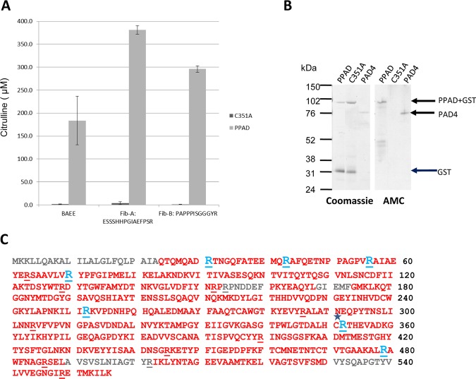
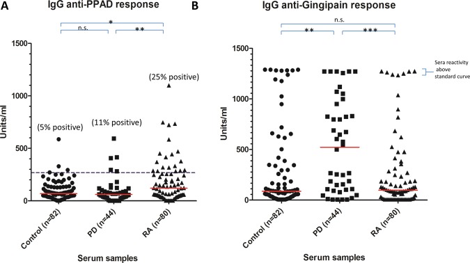
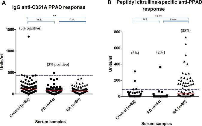
Results: Recombinant PPAD was a potent citrullinating enzyme. Antibodies to PPAD, but not to Rgp, were elevated in the RA sera (median 122 U/ml) compared with controls (median 70 U/ml; p<0.05) and PD (median 60 U/ml; p<0.01). Specificity of the anti-peptidyl citrullinated PPAD response was confirmed by the reaction of RA sera with multiple epitopes tested with synthetic citrullinated peptides spanning the PPAD molecule. The elevated antibody response to PPAD was abolished in RA sera if the C351A mutant was used on ELISA.

Conclusions: The peptidyl citrulline-specific immune response to PPAD supports the hypothesis that, as a bacterial protein, it might break tolerance in RA, and could be a target for therapy.

Keywords: Ant-CCP; Autoimmunity; Rheumatoid Arthritis.


Figures

Figure 1
Figure 1
PPAD efficiently citrullinates BAEE and fibrinogen peptides and is autocitrullinated. (A) 1 µM C351A PPAD mutant and recombinant PPAD were tested for enzyme activity using 1 mM BAEE, Fib-A or Fib-B as substrate, with citrullinated product quantified spectrometrically. (B) Equivalent amounts of recombinant GST-His-PPAD (PPAD, 89 kDa), inactive mutant (C351A, 89 kDa) and PAD4 (76 kDa) were resolved by SDS-PAGE and Coomassie stained. The resolved proteins were transferred onto nitrocellulose membranes and protein citrullination detected with an antimodified citrulline antibody. (C) Mass spectrometry sequence coverage map of PPAD. Sequence in red indicates the identified sequence. Arginine residues are underlined. Arginine residues that were modified to citrulline are shown in blue. C* indicates the Cysteine residue mutated to produce C351A inactive mutant.
Figure 2
Figure 2
PPAD associates with the bacterial cell and is expressed on the outer membrane (OM). (A) An immortalised oral keratinocyte cell line (OKF6) was stained with anti-PPAD antibody (i). Cells were infected with Porphyromonas gingivalis and stained with anti-PPAD antibody (ii) or isotype control (iii). Cell nuclei were counterstained blue. Magnification: ×400. (B) PPAD subcellular localisation in P gingivalis. Bacteria culture from an early stationary phase was fractionated into whole cell extract (WCE), particle-free growth media (Media), soluble cell proteins derived from periplasm and cytoplasm (PP/CP), cell envelope containing inner and outer membranes (CE), inner membrane (IM) and outer membrane (OM). Fractions were subjected to Western blot analysis using anti-PPAD antibody.
Figure 3
Figure 3
ELISA analysis of (A) anti-PPAD and (B) anti-RgpB antibodies in controls, periodontitis and rheumatoid arthritis serum. The red line indicates median units per ml. Mann–Whitney U test was used to calculate p values for differences between the groups (n.s.=no significant difference, *=p<0.05 and **=p<0.01, ***=p<0.001). The cut-off for anti-PPAD positive sera was calculated based on the 95th percentile of the control sample values (blue dashed line).
Figure 4
Figure 4
Peptidyl citrulline-specific antibody response. (A) Anti-C351A PPAD and (B) peptidyl citrulline specific anti-PPAD antibodies in controls, periodontitis and rheumatoid arthritis serum. The red line indicates median units per ml. Mann–Whitney U test was used to calculate p values for differences between the groups (n.s.=no significant difference, *=p<0.05 and **=p<0.01, ***=p<0.001, ****=p<0.0001). The units/ml of the anti-C351A PPAD response were subtracted from the units/ml of the anti-PPAD response giving the specific level of anti-peptidyl citrulline antibody response. The cut-off for the peptidyl citrulline specific anti-PPAD antibodies was calculated based on the 95th percentile of the control sample values (blue dashed line).
Figure 5
Figure 5
Correlation of antibody detection by ELISA and immunoblotting. Representative immunoblot positive and negative of serum reacting to PPAD (PPAD-GST 89 kDa) and RgpB (RgpB-His 47kDa) (A). Validation of the ELISA analysis relative to the immunoblotting results (B+C). The 27 sera (10 control, 7 peridontitis and 10 rheumatoid arthritis) were divided into two groups based on presence (+) or absence (−) of IgG antibodies to (B) PPAD or (C) RgpB as determined by immunoblotting. The red line indicates the ELISA mean U/ml value for the serum groups. The Mann–Whitney U test was used to calculate p values for the difference between the arbitury units for the anti-PPAD/anti-RgpB blot positive (+) serum group and blot negative (−) serum group (** p<0.05 and ***p<0.01).

References

    1. Wegner N, Lundberg K, Kinloch A, et al. Autoimmunity to specific citrullinated proteins gives the first clues to the etiology of rheumatoid arthritis. Immunol Rev 2010;233:34–54 - PubMed
    1. Vossenaar ER, Zendman AJ, van Venrooij WJ, et al. PAD, a growing family of citrullinating enzymes: genes, features and involvement in disease. Bioessays 2003;25:1106–18 - PubMed
    1. Vossenaar ER, Smeets TJ, Kraan MC, et al. The presence of citrullinated proteins is not specific for rheumatoid synovial tissue. Arthritis Rheum 2004;50:3485–94 - PubMed
    1. Foulquier C, Sebbag M, Clavel C, et al. Peptidyl arginine deiminase type 2 (PAD-2) and PAD-4 but not PAD-1, PAD-3, and PAD-6 are expressed in rheumatoid arthritis synovium in close association with tissue inflammation. Arthritis Rheum 2007;56:3541–53 - PubMed
    1. Kinloch A, Lundberg K, Wait R, et al. Synovial fluid is a site of citrullination of autoantigens in inflammatory arthritis. Arthritis Rheum 2008;58:2287–95 - PubMed

Publication types

MeSH terms