. 2013 Apr;44(4):1158-61.
doi: 10.1161/STROKEAHA.111.000033. Epub 2013 Mar 5.

Quantitative video-oculography to help diagnose stroke in acute vertigo and dizziness: toward an ECG for the eyes

Affiliations

Quantitative video-oculography to help diagnose stroke in acute vertigo and dizziness: toward an ECG for the eyes

David E Newman-Toker et al. Stroke. 2013 Apr.

Abstract

Background and purpose: Strokes can be distinguished from benign peripheral causes of acute vestibular syndrome using bedside oculomotor tests (head impulse test, nystagmus, test-of-skew). Using head impulse test, nystagmus, test-of-skew is more sensitive and less costly than early magnetic resonance imaging for stroke diagnosis in acute vestibular syndrome but requires expertise not routinely available in emergency departments. We sought to begin standardizing the head impulse test, nystagmus, test-of-skew diagnostic approach for eventual emergency department use through the novel application of a portable video-oculography device measuring vestibular physiology in real time. This approach is conceptually similar to ECG to diagnose acute cardiac ischemia.

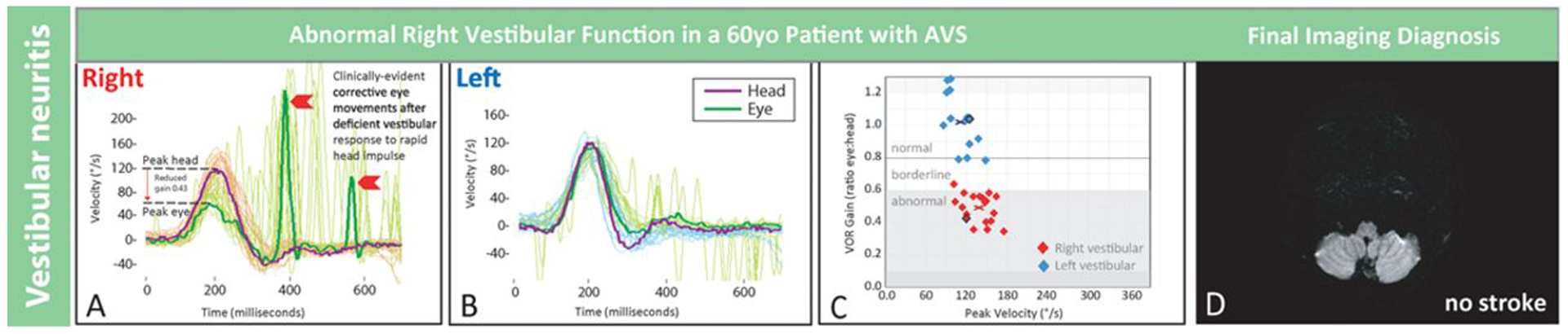
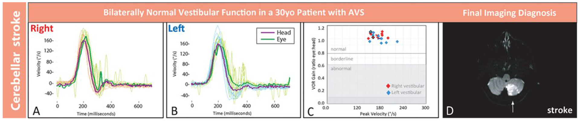
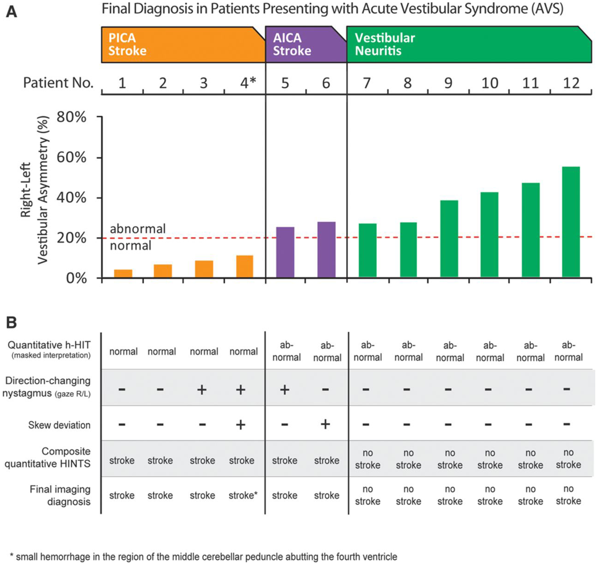
Methods: Proof-of-concept study (August 2011 to June 2012). We recruited adult emergency department patients with acute vestibular syndrome defined as new, persistent vertigo/dizziness, nystagmus, and (1) nausea/vomiting, (2) head motion intolerance, or (3) new gait unsteadiness. We recorded eye movements, including quantitative horizontal head impulse testing of vestibulo-ocular-reflex function. Two masked vestibular experts rated vestibular findings, which were compared with final radiographic gold-standard diagnoses. Masked neuroimaging raters determined stroke or no stroke using magnetic resonance imaging of the brain with diffusion-weighted imaging obtained 48 hours to 7 days after symptom onset.

Results: We enrolled 12 consecutive patients who underwent confirmatory magnetic resonance imaging. Mean age was 61 years (range 30-73), and 10 were men. Expert-rated video-oculography-based head impulse test, nystagmus, test-of-skew examination was 100% accurate (6 strokes, 6 peripheral vestibular).

Conclusions: Device-based physiological diagnosis of vertebrobasilar stroke in acute vestibular syndrome should soon be possible. If confirmed in a larger sample, this bedside eye ECG approach could eventually help fulfill a critical need for timely, accurate, efficient diagnosis in emergency department patients with vertigo or dizziness who are at high risk for stroke.


Figures

Figure 1.
Figure 1.
Quantitative head impulse and magnetic resonance imaging (MRI) results in an older patient with vestibular neuritis. Shown are results from patient No. 8 in Figure 3, a 60-year-old man with vestibular neuritis. Clinical presenting features were vertigo, nausea, and gait disturbance without general neurological or auditory symptoms or signs. There was unidirectional nystagmus and no skew deviation. Shown are physiological tracings from multiple rightward (A) and leftward (B) horizontal head impulse test (h-HIT) maneuvers temporally superimposed with a single maneuver highlighted darker. The rightward h-HIT response is abnormal (A). It has both a quantitative abnormality (reduced vestibulo-ocular reflex [VOR] gain during the head movement, downward red arrow) and a qualitative, clinically evident abnormality (fast, corrective eye movements to realign the eye on the target after the head stops moving, red chevrons). The leftward h-HIT response is normal (B). Each h-HIT result is mapped in the corresponding VOR gain plot (C), where the central x denotes the mean right- (red) or left-sided (blue) VOR gain across h-HIT trials. A single representative axial MRI-diffusion-weighted images (DWI) through the inferior cerebellum shows no stroke (D). Note that only the abnormal h-HIT result differed from clinical findings in patient No. 2, the young patient with a large, inferior cerebellar stroke in Figure 2. AVS indicates acute vestibular syndrome and °/s, degrees per second.
Figure 2.
Figure 2.
Quantitative head impulse and magnetic resonance imaging (MRI) results in a young patient with stroke. Shown are results from patient No. 2 in Figure 3, a 30-year-old man with inferior cerebellar stroke. Clinical presenting features were vertigo, nausea, and gait disturbance without general neurological or auditory symptoms or signs. There was unidirectional nystagmus and no skew deviation. Shown are physiological tracings from multiple rightward (A) and leftward (B) horizontal head impulse test (h-HIT) maneuvers temporally superimposed with a single maneuver highlighted darker. Tracings reveal normal h-HIT results bilaterally, with a vestibulo-ocular reflex (VOR) eye movement response that closely matches the head movement (A and B). Each h-HIT result is mapped in the corresponding VOR gain plot (C), where the central x denotes the mean right- (red) or left-sided (blue) VOR gain across h-HIT trials. A single representative axial MRI–diffusion-weighted images (DWI) through the inferior cerebellum shows a large acute, left posterior inferior cerebellar artery territory infarction (D, arrow). The stroke was extensive (maximal dimensions 3.0×5.0×4.4 centimeters), spanning 8 axial sections. Note that only the bilaterally normal h-HIT result differed from clinical findings in patient No. 8, the older patient with vestibular neuritis in Figure 1. AVS indicates acute vestibular syndrome; and °/s, degrees per second.
Figure 3.
Figure 3.
Vestibulo-ocular reflex (VOR) abnormalities and oculographic head impulse, nystagmus, test-of-skew (HINTS) results for 12 acute vestibular syndrome (AVS) patients. A, Relative right-left VOR asymmetry for each patient (absolute VOR gains shown in Figure II in the online-only Data Supplement). Patients are grouped by diagnosis and arrayed from least to greatest asymmetry within groups. B, Masked interpretations of all quantitative and clinical results as well as composite HINTS-based diagnosis. Bilaterally normal horizontal head impulse test (h-HIT) results or those with direction-changing nystagmus or skew-predicted radiographic stroke. Unilaterally abnormal h-HIT results indicated vestibular neuritis, absent direction-changing nystagmus, and skew. AICA indicates anterior inferior cerebellar artery; PICA, posterior inferior cerebellar artery; and R/L, right/left.

Similar articles

Cited by

References

    1. Newman-Toker DE, Hsieh YH, Camargo CA, Pelletier AJ, Butchy GT, Edlow JA. Spectrum of dizziness visits to US emergency departments: cross-sectional analysis from a nationally representative sample. Mayo Clin Proc. 2008;83:765–775. - PMC - PubMed
    1. Tarnutzer AA, Berkowitz AL, Robinson KA, Hsieh YH, Newman-Toker DE. Does my dizzy patient have a stroke? A systematic review of bedside diagnosis in acute vestibular syndrome. CMAJ. 2011;183:E571–E592. - PMC - PubMed
    1. Newman-Toker DE, Kattah JC, Alvernia JE, Wang DZ. Normal head impulse test differentiates acute cerebellar strokes from vestibular neuritis. Neurology. 2008;70(24 pt 2):2378–2385. - PubMed
    1. Kattah JC, Talkad AV, Wang DZ, Hsieh YH, Newman-Toker DE. HINTS to diagnose stroke in the acute vestibular syndrome: three-step bedside oculomotor examination more sensitive than early MRI diffusion-weighted imaging. Stroke. 2009;40:3504–3510. - PMC - PubMed
    1. Chalela JA, Kidwell CS, Nentwich LM, Luby M, Butman JA, Demchuk AM, et al.Magnetic resonance imaging and computed tomography in emergency assessment of patients with suspected acute stroke: a prospective comparison. Lancet. 2007;369:293–298. - PMC - PubMed

Publication types