. 2013 Apr 23;7(4):3264-75.
doi: 10.1021/nn305958y. Epub 2013 Mar 20.

Targeted intraceptor nanoparticle therapy reduces angiogenesis and fibrosis in primate and murine macular degeneration

Affiliations

Targeted intraceptor nanoparticle therapy reduces angiogenesis and fibrosis in primate and murine macular degeneration

Ling Luo et al. ACS Nano. .

Abstract

Monthly intraocular injections are widely used to deliver protein-based drugs that cannot cross the blood-retina barrier for the treatment of leading blinding diseases such as age-related macular degeneration (AMD). This invasive treatment carries significant risks, including bleeding, pain, infection, and retinal detachment. Further, current therapies are associated with a rate of retinal fibrosis and geographic atrophy significantly higher than that which occurs in the described natural history of AMD. A novel therapeutic strategy which improves outcomes in a less invasive manner, reduces risk, and provides long-term inhibition of angiogenesis and fibrosis is a felt medical need. Here we show that a single intravenous injection of targeted, biodegradable nanoparticles delivering a recombinant Flt23k intraceptor plasmid homes to neovascular lesions in the retina and regresses CNV in primate and murine AMD models. Moreover, this treatment suppressed subretinal fibrosis, which is currently not addressed by clinical therapies. Murine vision, as tested by OptoMotry, significantly improved with nearly 40% restoration of visual loss induced by CNV. We found no evidence of ocular or systemic toxicity from nanoparticle treatment. These findings offer a nanoparticle-based platform for targeted, vitreous-sparing, extended-release, nonviral gene therapy.


Figures
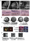
Fig. 1
Fig. 1. Knockdown (AAV.shRNA.sFlt-1) induces murine CNV
(A) Secondary CNV was observed late and distant from injection site on fluorescein angiography (FA, top). Secondary CNV was detected at 10 weeks at a location where CNV was not observed by imaging at 6 weeks after injection and was distant from primary CNV at the injection site. H&E staining (bottom) shows the histology of normal retina, secondary CNV and classic CNV from the injection site in the sFlt-1 knockdown mouse (6 months after injection), respectively (40×). (B) Classic CNV, subretinal fluid and intrachoroidal CNV or occult CNV were detected in the same eye (at 6 months).(PED: Pigment epithelial detachment; the top row images are Indocyanine Green ( ICG)Angiography images.(C) IHC staining on choroidal flat mounts from sFlt-1 knockdown eyes (6 months after injection) showed neovessels (isolectin) and fibrosis (collagen I) in CNV site. (D) The morphology of sFlt-1 knockdown induced CNV closely mimics human CNV (from the database of Heidelberg Spectralis imager), while the retina in laser-induced CNV eyes displays partial burnout (arrow). (E) IHC staining demonstrates that alpha5 integrin was expressed strongly in all three types of CNV models (scale bar: 100 μm).
Fig. 2
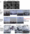
Fig. 2. RGD-functionalized nanoparticles localize to CNV lesions
(A) RGD.Flt23k.NR.NP was detected by the Spectralis imager in vivo in CNV eyes and normal eyes. (B) On histological confocal examination of murine ocular cryosections, more nanoparticles are present in eyes with CNV than in normal eyes. Nanoparticles were concentrated in CNV lesions at 24 hours post-intravenous injection. RGD-conjugated nanoparticles were detected in CNV lesions 14 days post-injection while only up to 7 days in normal eyes. (a, b) Magnified images from the frames show nanoparticles in CNV site and normal site in the same eye. (C) In laser-induced CNV monkey eyes one month post-intervention, some RGD-conjugated nanoparticles could still be detected in CNV lesion. However, unconjugated or blank nanoparticles were not found in CNV lesions.
Fig. 3
Fig. 3. Angiogenesis is inhibited in all CNV models, accompanied by restored vision in CNV eyes induced by sFlt-1 knockdown
(A) In the laser-induced CNV monkey model, the volumes of CNV lesions, including neovessels (stained by perlecan) and fibrosis (stained by collagen I), significantly decreased 4 weeks after RGD.Flt23k.NR.NP treatment (a). Representative images of FA (b), IHC staining(c) and H&E staining (d)show the different size of CNV in each group. (B) In laser-induced CNV mice, CNV lesions were regressed byRGD.Flt23k.NR.NPtwo weeks post-intervention. (C) In the sFlt-1 knockdowninduced murine CNV model, the volumes of CNV lesions were significantly decreased by RGD.Flt23k.NR.NP at 4 weeks after treatment. (D) Representative FA and OCT images detected the decreased CNV in treated mice from Fig.3C, and detected increased CNV accompanied with secondary CNV occurrence in controls (arrows point to CNV). (E) Representative H&E images of each group in Fig. 3C show the different sizes of CNV after intervention. (F) Single systemic administration of RGD.Flt23k.NR.NP regressed CNV more than intravitreal anti-VEGF antibody. (G) Optomotor testingvision function was partially restored after 4 weeks treatment with RGD.Flt23k.NR.NP but not with the vehicle or sham controls.
Fig. 4
Fig. 4. Ocular and systemic toxicity were not detected after RGD.Flt23k.NR.NP treatment
Representative monkey ocular fundus images 30 days after intervention showed no significant retinal morphological difference between treatment and control. (B) No apoptosis in the retina outside the CNV lesions was detected at day 30 post intervention by TUNEL staining (Scale bar: 100μm). (C) H&E images showed no significant histopathological changes in the liver, lung, kidney, and skin sections after intravenous delivery of nanoparticles in comparison to untreated, normal organs (30 days).

References

    1. Ambati J, Ambati BK, Yoo SH, Ianchulev S, Adamis AP. Age-Related Macular Degeneration: Etiology, Pathogenesis, and Therapeutic Strategies. Surv Ophthalmol. 2003:257–293. - PubMed
    1. Bunce C, Xing W, Wormald R. Causes of Blind and Partial Sight Certifications in England and Wales: April 2007–March 2008. Eye (Lond) 2010:1692–1699. - PubMed
    1. Jager RD, Mieler WF, Miller JW. Age-Related Macular Degeneration. N Engl J Med. 2008:2606–2617. - PubMed
    1. Pascolini D, Mariotti SP, Pokharel GP, Pararajasegaram R, Etya'ale D, Negrel AD, Resnikoff S. 2002 Global Update of Available Data on Visual Impairment: A Compilation of Population-Based Prevalence Studies. Ophthalmic Epidemiol. 2004:67–115. - PubMed
    1. Alon T, Hemo I, Itin A, Pe'er J, Stone J, Keshet E. Vascular Endothelial Growth Factor Acts as a Survival Factor for Newly Formed Retinal Vessels and Has Implications for Retinopathy of Prematurity. Nat Med. 1995:1024–1028. - PubMed

Publication types