Review
. 2013 May;38(5):233-42.
doi: 10.1016/j.tibs.2013.01.004. Epub 2013 Mar 1.

Nutrient signaling to mTOR and cell growth

Affiliations
Review

Nutrient signaling to mTOR and cell growth

Jenna L Jewell et al. Trends Biochem Sci. 2013 May.

Abstract

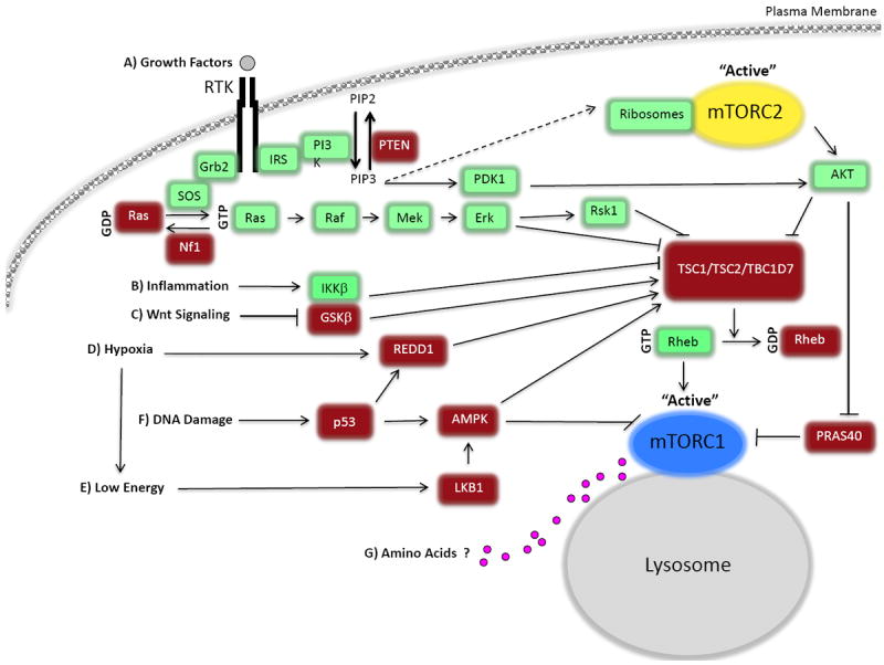
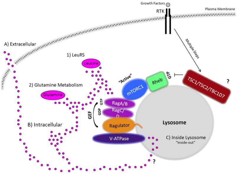
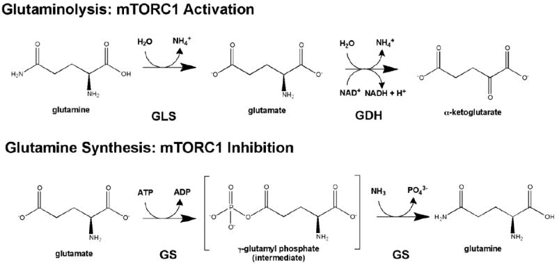
The mammalian target of rapamycin (mTOR) is a conserved protein kinase involved in a multitude of cellular processes including cell growth. Increased mTOR activation is observed in multiple human cancers and inhibition of mTOR has proven efficacious in numerous clinical trials. mTOR comprises two complexes, termed mTORC1 and mTORC2. Both complexes respond to growth factors, whereas only mTORC1 is controlled by nutrients, such as glucose and amino acids. Since the discovery of mTOR, extensive studies have intricately detailed the molecular mechanisms by which mTORC1 is regulated. Somewhat paradoxically, amino acid (AA)-induced mTORC1 activation -arguably the most essential stimulus leading to mTORC1 activation - is the least understood. Here we review the current knowledge of nutrient-dependent regulation of mTORC1.


Figures

Figure 1
Figure 1. The mTOR signaling cascade
Mammalian target of rapamycin complex 1 (mTORC1) and 2 (mTORC2) are both controlled by growth factors, whereas mTORC1 is also regulated by cellular energy status, oxygen, stress, and amino acids (AAs). A) mTOR is controlled by growth factors through the classical phosphatidylinositide 3-kinase-protein kinase B (also known as AKT), PI3K-AKT, pathway and through the Ras signaling cascade. PI3K is activated and recruited to the membrane by insulin receptor substrate (IRS-1), where it catalyzes the phosphorylation of PtdIns(4,5)P2 (PIP2) to PtdIns(3,4,5)P3 (PIP3). AKT is recruited via its pleckstrin homology (PH) domain binding to PIP3, and is phosphorylated and activated by the phosphoinositide-dependent kinase-1 (PDK1). mTORC2 is thought to be activated downstream of PI3K, possibly by ribosome binding, although many unanswered questions still remain. Dashed arrow represents missing components possibly involved that have not been identified. Full activation of AKT requires the phosphorylation of T308 (by PDK1) and S473 (by mTORC2). Although, the requirement of AKT phosphorylation at T308 or S473 depends on its substrate. AKT kinase activation can promote mTORC1 signaling in two ways: by phosphorylating and inhibiting tuberous sclerosis complex 2 (TSC2) GTPase activating protein (GAP) activity, thus activating Rheb, and by phosphorylating proline-rich AKT/PKB substrate 40 kDa (PRAS40), which increases PRAS40-14-3-3 binding and relieves PRAS40 inhibition on mTORC1. In addition to AKT, the Ras pathway regulates mTORC1 through ERK and RSK1, which phosphorylate and inhibit TSC1/2. TBC1D7, a third subunit of TSC, promotes TSC1-TSC2 interaction and Rheb-GAP activity. RTK, denotes receptor tyrosine kinase. B) IKKβ can also inhibit TSC1/TSC2 in response to inflammation. C) GSK3-β, which is regulated by Wnt signaling, phosphorylates TSC2 and increases its GAP activity in an AMP-activated dependent kinase (AMPK)-priming phosphorylating manner. D) Hypoxia promotes expression of the regulated in development and DNA damage responses 1 (REDD1), which activates TSC1/TSC2, and hypoxia activates AMPK due to the failure to generate sufficient ATP. E) When cellular energy is low AMPK is activated and regulates mTORC1 by phosphorylating and activating TSC2. Also, AMPK can phosphorylate Raptor and inhibit mTORC1 function. F) DNA damage results in the inhibition of mTORC1 activity through the p53-dependent up-regulation of REDD1 and AMPK. G) AA signaling activates mTORC1 (see Figure 3). Pink circles represent AAs.
Figure 2
Figure 2. Energy sensing by AMPK and control of mTORC1
AMP-activated protein kinase (AMPK) acts as a metabolic checkpoint under nutrient starvation conditions, translating signals to mTORC1 through direct phosphorylation of tuberous sclerosis complex 2 (TSC2) and Raptor and inhibiting cell growth. The phosphorylation of AMPK at threonine 172 by liver kinase B1 (LKB1) in the activation loop is essential for AMPK activation. LKB1 is in a complex with STRAD and MO25. AMPK phosphorylates and activates TSC1/2 by phosphorylating TSC2, which inhibits mTORC1. AMPK also phosphorylates regulatory-associated protein of mTOR (Raptor) in a parallel pathway on sites that induce Raptor-14-3-3 binding and inhibition of mTORC1. RTK denotes receptor tyrosine kinase. P represents phosphorylation.
Figure 3
Figure 3. Amino acid signaling to mTORC1
Amino acids (AAs), depicted as pink circles, may be sensed A) extracellularly, B) intracellularly, from within the C) lysosome, or through a combination. In addition, the specific AA sensed to promote mTORC1 activation is still not clear. For example, mTORC1 can sense 1) leucine through leucyl-tRNA synthetase (LeuRS) and 2) glutamine levels. Glutaminolysis results in mTORC1 activation, whereas glutamine synthesis seems to inhibit mTORC1 (Box 1). In response to AAs, an active RagA/BGTP-RagC/DGDP physically interacts with regulatory-associated protein of mTOR (Raptor) and recruits mTORC1 to the lysosome to promote its activation by Rheb, another GTPase. Recently, TSC2 has also been reported to localize at the lysosome (TSC1 and TBC1D7 have not yet been identified to reside at the lysosome, denoted by ?). The mTORC1 interaction with Rag A/BGTP-Rag C/DGDP is anchored to the lysosome by a complex called the Ragulator. The Ragulator also serves as a guanine exchange factor (GEF) for Rag A/BGTP, promoting mTORC1 activation. Moreover, the v-ATPase has been observed to be upstream of the Ragulator in AA sensing to mTORC1. The v-ATPase senses AA from within the lysosomal lumen, through an “inside-out” type of mechanism. How AAs accumulate inside the lysosome and what transporter(s) are involved in this are still unknown, denoted by ?. Growth factors, through the PI3K-AKT-TSC pathway, activate Rheb (RhebGTP) so that it can turn on the kinase activity of mTORC1, whereas AAs through the Rags localize mTORC1 in close proximately to Rheb at the lysosome. RTK denotes receptor tyrosine kinase.
Figure I
Figure I. mTORC1 activation controlled by glutamine
Glutamine metabolism controls mTORC1. Top- Glutaminolysis, the metabolism of glutamine to α-ketoglutarate (αKG), is a two step process which results in the actiavtion of mTORC1. Glutamine is first de-aminated by glutaminase (GLS) to produce glutamate, and then glutamate dehydrogenase (GDH) produces a-KG. Bottom- In contrast, the synthesis of glutamine inhibits mTORC1 activation. Glutamine is synthesized by glutamine synthetase from glutamate.

References

    1. Heitman J, et al. Targets for cell cycle arrest by the immunosuppressant rapamycin in yeast. Science. 1991;253:905–909. - PubMed
    1. Kunz J, et al. Target of rapamycin in yeast, TOR2, is an essential phosphatidylinositol kinase homolog required for G1 progression. Cell. 1993;73:585–596. - PubMed
    1. Sabatini DM, et al. RAFT1: a mammalian protein that binds to FKBP12 in a rapamycin-dependent fashion and is homologous to yeast TORs. Cell. 1994;78:35–43. - PubMed
    1. Sabers CJ, et al. Isolation of a protein target of the FKBP12-rapamycin complex in mammalian cells. J Biol Chem. 1995;270:815–822. - PubMed
    1. Brown EJ, et al. A mammalian protein targeted by G1-arresting rapamycin-receptor complex. Nature. 1994;369:756–758. - PubMed

Publication types

MeSH terms

Substances

LinkOut - more resources