. 2013 Jul:60:282-91.
doi: 10.1016/j.freeradbiomed.2013.02.029. Epub 2013 Mar 4.

Induction of oxidative and nitrosative damage leads to cerebrovascular inflammation in an animal model of mild traumatic brain injury induced by primary blast

Affiliations

Induction of oxidative and nitrosative damage leads to cerebrovascular inflammation in an animal model of mild traumatic brain injury induced by primary blast

P M Abdul Muneer et al. Free Radic Biol Med. 2013 Jul.

Abstract

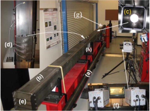
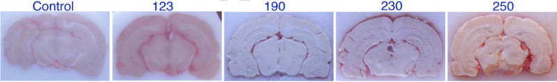
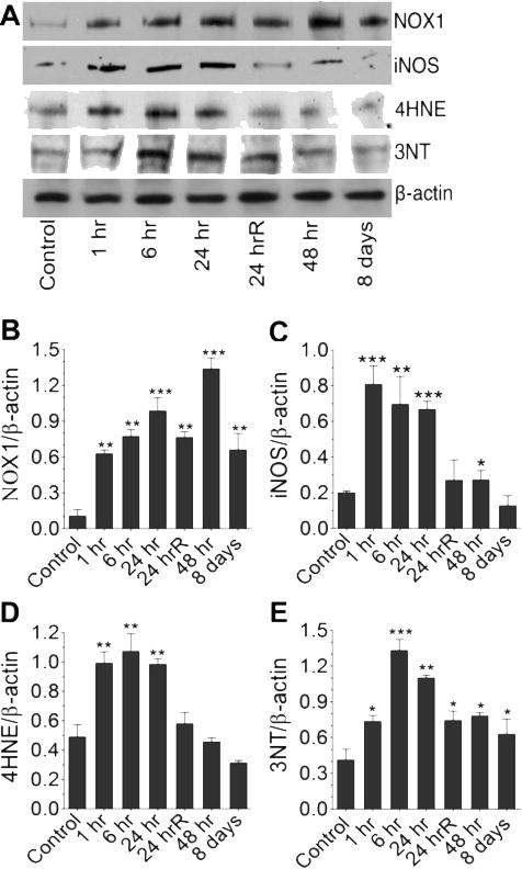
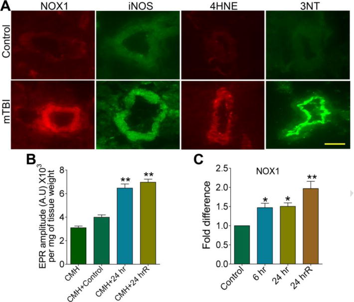
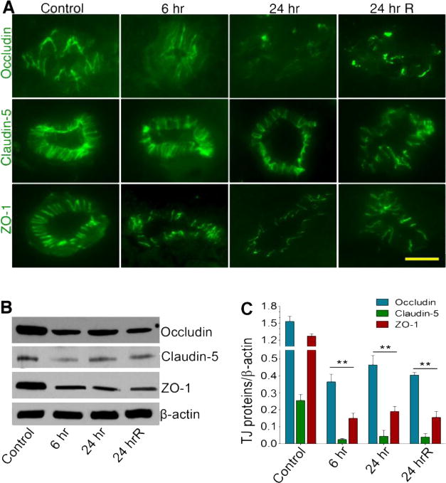
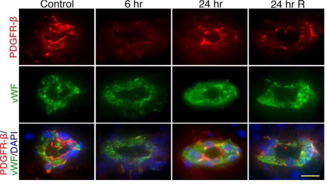
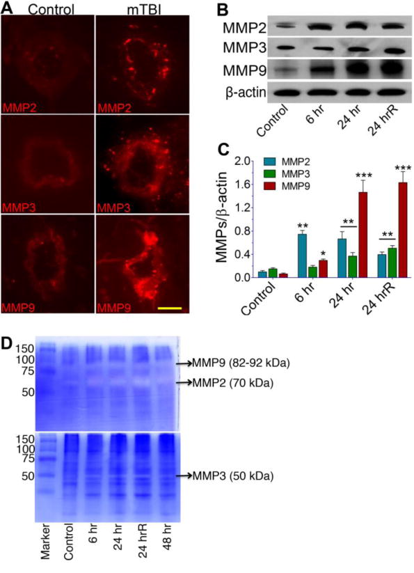
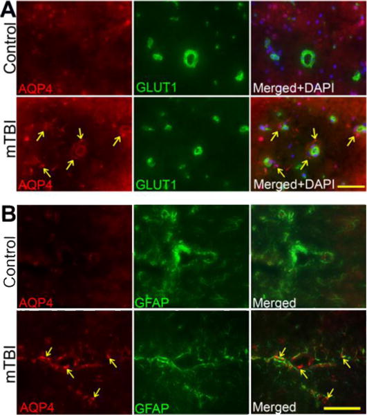
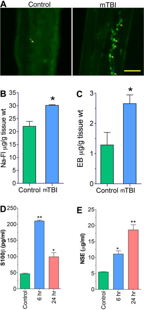
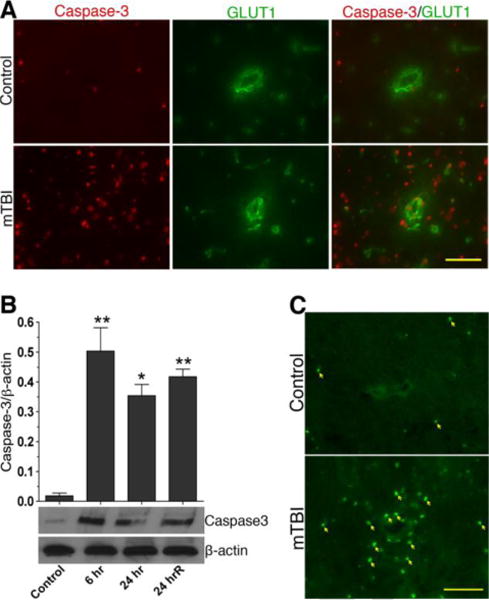
We investigate the hypothesis that oxidative damage of the cerebral vascular barrier interface (the blood-brain barrier, BBB) causes the development of mild traumatic brain injury (TBI) during a primary blast-wave spectrum. The underlying biochemical and cellular mechanisms of this vascular layer-structure injury are examined in a novel animal model of shock tube. We first established that low-frequency (123kPa) single or repeated shock wave causes BBB/brain injury through biochemical activation by an acute mechanical force that occurs 6-24h after the exposure. This biochemical damage of the cerebral vasculature is initiated by the induction of the free radical-generating enzymes NADPH oxidase 1 and inducible nitric oxide synthase. Induction of these enzymes by shock-wave exposure paralleled the signatures of oxidative and nitrosative damage (4-HNE/3-NT) and reduction of the BBB tight-junction (TJ) proteins occludin, claudin-5, and zonula occluden 1 in the brain microvessels. In parallel with TJ protein disruption, the perivascular unit was significantly diminished by single or repeated shock-wave exposure coinciding with the kinetic profile. Loosening of the vasculature and perivascular unit was mediated by oxidative stress-induced activation of matrix metalloproteinases and fluid channel aquaporin-4, promoting vascular fluid cavitation/edema, enhanced leakiness of the BBB, and progression of neuroinflammation. The BBB leakiness and neuroinflammation were functionally demonstrated in an in vivo model by enhanced permeativity of Evans blue and sodium fluorescein low-molecular-weight tracers and the infiltration of immune cells across the BBB. The detection of brain cell proteins neuron-specific enolase and S100β in the blood samples validated the neuroastroglial injury in shock-wave TBI. Our hypothesis that cerebral vascular injury occurs before the development of neurological disorders in mild TBI was further confirmed by the activation of caspase-3 and cell apoptosis mostly around the perivascular region. Thus, induction of oxidative stress and activation of matrix metalloproteinases by shock wave underlie the mechanisms of cerebral vascular BBB leakage and neuroinflammation.


Figures

Figure 1
Figure 1. Blast wave simulation and testing facility at University of Nebraska, Lincoln
(a) 4″ cylindrical shock tube; (a) 9″ square shock tube; (c) transition section; (d) pressure sensor array; (e) adjustable end reflector; and (f) Photron SA1.1 high speed cameras, (g) driver section (h) test section.
Figure 2
Figure 2. Mechanical cerebrovascular injury by different blasts exposure
The figure shows neurovascular or brain damage due to different blast exposure of mTBI at 123, 190, 230 and 250 kPa after 24 hr of post-exposure.
Figure 3
Figure 3. mTBI induces free radical adducts in rat brain
(A) Western blot analyses of NOX1, iNOS, 4HNE and 3NT in the whole rat brain homogenates at different time points after exposure to blast with 123 kPa peak. (B–E) Bar graphs show the results that are expressed as ratio of NOX1/iNOS/4HNE/3NT to that of β-actin band. Values are mean ±SEM (n = 4). *p<0.05; **p<0.01, ***p<0.001 versus control.
Figure 4
Figure 4. Primary blast induced oxidative and nitrosative stress in the rat brain microvessels
(A) Immunofluorescent staining of NOX1, iNOS, 4HNE, and 3NT in intact brain microvessels of rats subjected to a single blast with 123 kPa peak overpressure, 24 hours post-exposure. (B) ROS generation was detected by electron paramagnetic resonance (EPR) in brain tissue slices from 24 hr and 24 hrR post-exposure of mTBI of 123 kPa blast exposed and compared with control. Results are expressed in EPR amplitude arbitrary units per milligrams of tissue weight. (C) Changes in mRNA level of NOX1 in brain cortical tissues of rats in different time intervals of 123 kPa blast post-exposure by quantitative RT-PCR using TaqMan primers. Values are mean ±SEM (n = 3 in B and C). Statistically significant *p<0.05; **p<0.01 versus control in C; and versus CMH+control in B. Scale bar: 5 μm in all panels of A.
Figure 5
Figure 5. Primary blast causes impairment in tight junction proteins
(A) Immunofluorescent staining of tight junction proteins: Occludin, Claudin-5 and ZO-1 in intact brain microvessels of rats at different time periods post-exposure (blast with 123 kPa peak overpressure): 6 hr, 24 hr and 24 hrR (two repeated exposures of every 24 hours). (B) Western blot analysis of Occludin, Claudin-5, ZO-1 and actin in the whole brain tissue homogenates of rats at different time points after exposure to blast (123 kPa peak overpressure). (C) Graph shows the results that are expressed as ratio of occludin/claudin5/ZO-1 to that of β-actin band. Values are mean ±SEM (n = 3). Statistically significant, **p<0.01 versus control in C. Scale bar:5 μm.
Figure 6
Figure 6. Pericytes have significant role in BBB dysfunction in primary blast-induced mTBI
Immunofluorescent staining of pericyte specific marker, PDGFR-β (red) and endothelial marker vWF (green) in intact brain microvessels of rats exposed to primary blast (123 kPa peak overpressure). Cell nuclei were counterstained with DAPI (blue). The expression of PDGFR-β decreased with time (6 and 24 hours, single exposure) and after repeated insult (24 hr R). Scale bar = 5 μm in all panels.
Figure 7
Figure 7. mTBI activates matrix metalloproteinases (MMPs)
(A) Immunofluorescent staining of MMP-2, MMP-3, and MMP-9 in intact brain microvessels of mTBI exposed and control rats. (B) Western blots analyses of MMP-2, MMP-3, MMP-9, and actin in whole brain tissue homogenates. (C) Graph shows the results that are expressed as ratio of MMP-2/3/9 to that of β-actin band. (D) The gelatinolytic activity of MMP-2/9 (top gel) and caseinolytic activity of MMP-3 (bottom gel) were demonstrated by gelatin or casein zymography in the rat brain cortical tissue protein extracts from different time periods such as 6 hr, 24 hr, 24 hrR and 48 hr of post-exposure of blast with 123 kPa peak overpressure. Values are mean ±SEM; n=4. Statistically significant, **p<0.01 ***p<0.001 versus control in C. Scale bar = 5 μm in all panels of A.
Figure 8
Figure 8. mTBI activates aquaporin-4
(A) Immunofluorescent staining of aquaporin-4 (AQP-4) (red) and endothelial marker, GLUT1 (green) in brain tissue sections contain microvessels of exposed to primary blast (123 kPa peak overpressure). Cell nuclei were counterstained with DAPI (blue). The arrow indicates the AQP-4 staining surrounding the microvessels. Scale bar = 40 μm in all panels. (B) Immunofluorescent staining of aquaporin-4 (AQP-4) (red) co-localized with GFAP (astrocyte marker-green) in brain tissue sections contain microvessels of exposed to primary blast (123 kPa peak overpressure). The arrow indicates the AQP-4 staining surrounding the microvessels. Scale bar = 40 μm in all panels.
Figure 9
Figure 9. mTBI causes BBB leakage
(A) Fluo-3 labeled macrophage adhesion/migration in brain capillary following infusion of cells into the common carotid artery of mTBI exposed rats after 24 hr of blast and compared with controls. (B,C) Graphical representation of In vivo permeability to show the leakage of Evans Blue (EB, 5 μM)(B) and Sodium fluorescein (Na-Fl, 5 μM) (C) in mTBI exposed rats. The carotid artery of rats was exposed and infused EB-NaFl mixture and collected the brain after one hour of infusion and processed for BBB permeability assay as given in materials and methods. (D, E) ELISA shows the levels of S100β (D) and NSE (E) in the blood serum of mTBI exposed rats. The blood serum was collected after 24 hr of blast exposure. Values are mean ±SEM, n=3 in B, C and n=5 in D,E. *p<0.05; **p<0.01 are statistically significant in BE. Scale bar =40 μm in both panels of A.
Figure 10
Figure 10. mTBI causes cell apoptosis
(A) Changes in the expression of active caspase-3 (red) in brain microvessels and cortical tissue sections of control and mTBI blast (123 kPa) exposed animals. (B) Immunoblot analysis to show the alterations in active caspase-3 protein levels in brain cortical tissue and microvessel homogenates proteins. Bar graphs show the results, which are expressed as ratio of caspase-3 (active) to that of β-actin bands. Values are mean ±SEM; n=4 to 5. *p<0.05; **p<0.01 are statistically significant. (C) TUNEL staining in rat brain cortical tissue section. The arrow indicates the TUNEL-positive cells. Scale bar = 20 μm in all panels of A and B.

References

    1. Hoge CW, McGurk D, Thomas JL, Cox AL, Engel CC, Castro CA. Mild traumatic brain injury in U.S. Soldiers returning from Iraq. N Engl J Med. 2008;358:453–463. - PubMed
    1. Vanderploeg RD, Belanger HG, Horner RD, Spehar AM, Powell-Cope G, Luther SL, Scott SG. Health Outcomes Associated With Military Deployment: Mild Traumatic Brain Injury, Blast, Trauma, and Combat Associations in the Florida National Guard. Arch Phys Med Rehabil. 2012 doi: 10.1016/j.apmr.2012.05.024. - DOI - PubMed
    1. Vasterling JJ, Verfaellie M, Sullivan KD. Mild traumatic brain injury and posttraumatic stress disorder in returning veterans: perspectives from cognitive neuroscience. Clin Psychol Rev. 2009;29:674–684. - PubMed
    1. Trudeau DL, Anderson J, Hansen LM, Shagalov DN, Schmoller J, Nugent S, Barton S. Findings of mild traumatic brain injury in combat veterans with PTSD and a history of blast concussion. J Neuropsychiatry Clin Neurosci. 1998;10:308–313. - PubMed
    1. Santiago PN, Wilk JE, Milliken CS, Castro CA, Engel CC, Hoge CW. Screening for alcohol misuse and alcohol-related behaviors among combat veterans. Psychiatr Serv. 2010;61:575–581. - PubMed

Publication types

MeSH terms

LinkOut - more resources