. 2013 May;10(3):396-404.
doi: 10.1016/j.scr.2013.01.007. Epub 2013 Jan 30.

Mammospheres from murine mammary stem cell-enriched basal cells: clonal characteristics and repopulating potential

Affiliations

Mammospheres from murine mammary stem cell-enriched basal cells: clonal characteristics and repopulating potential

Qiaoxiang Dong et al. Stem Cell Res. 2013 May.

Abstract

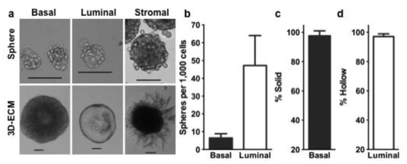
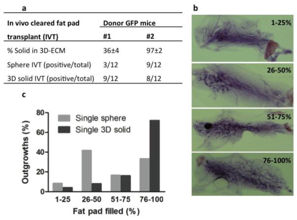
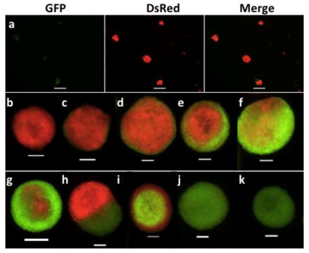
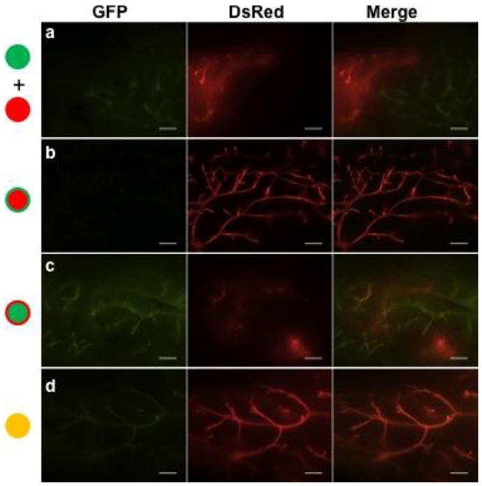
Identification of murine mammary stem cells (MaSCs) has been attempted with various in vitro and in vivo assays. While, the in vivo repopulation assay remains as the most definitive assay for MaSC detection, it is expensive, time-consuming, and technically challenging. The in vitro mammosphere assay was considered unreliable because of major concerns about its clonal origin. In the current study, co-culture experiments with mammary cells from fluorescent protein transgenic mice and time-lapse video microscopy revealed that >90% mammospheres formed from sorted basal epithelial-enriched cells were of clonal origin in terms of stem cell. These basal-cell derived mammospheres were further distinguished morphologically in a 3-dimensional extracellular matrix culture and functionally in the in vivo repopulation assay. Transplant of single mammospheres or the resultant 3-dimensional solid structures into gland-free mammary fat pads yielded a 70% success rate of multilineage mammary gland reconstitution. Thus, this in vitro sphere formation and differentiation assay is a reliable alternative to the in vivo repopulation assay for the study of MaSCs.


Figures

Figure 1
Figure 1
Characteristics of mammospheres derived from different fractions of fluorescence-activated cell sorting (FACS) sorted cells from wild type C57BL/6 mice (a). Single spheres were further plated in Matrigel to form corresponding morphologically unique 3-dimentional extracellular matrix (3D-ECM) structures (a). Bar graphs showing the sphere formation efficiency (b) and percentage of solid (c) or hollow (d) structures in 3D-ECM culture. Scale bars, 100 μm. The mean and standard deviation values, and the number of replicates used for each plotted figures are stated in the Results.
Figure 2
Figure 2
In vivo cleared mammary fat pad transplant of single mammosphere and single 3D-ECM solid structure derived from FACS sorted basal cell fraction (CD24+CD49fhi) of two transgenic green fluorescent protein (GFP) mice (a). Representative whole mount staining pictures showing different percentage of fill in cleared fat pad with regenerated GFP glands (b). Bar graphs showing percentage of fill in cleared fat pad for all regenerated glands derived from single mammosphere (light gray bars) or single 3D-ECM solid structure (dark gray bars) with in vivo transplant (c).
Figure 3
Figure 3
Mammospheres (a) derived from co-culture of FACS sorted basal cells of GFP and DsRed mice, and subsequent 3D-ECM culture derived solid structures (b–k). Scale bars, 100 μm.
Figure 4
Figure 4
Regenerated glands from in vivo transplant of (a) one 3D solid structure from DsRed mice together with one 3D solid structure from GFP mice or of (b–d) single 3D structures derived from spheres formed from co-culture of FACS sorted basal cells of GFP and DsRed mice that were characterized by predominantly red (b), predominantly green (c), and true chimeras (d). Scale bars, 500 μm.
Figure 5
Figure 5
Percent basal cell gated within the LinCD24+CD49fhi fraction (a), total number of basal cells per mouse (b), %SFD-IC in total epithelial cell (c) and fold increase (d) in basal SFD-IC number from pairs of age-matched virgin (light gray bars) and 12.5-day pregnant (dark gray bars) FVB (n = 4 pairs of same age, approximately 2.5-month old) or C57BL/6 (n = 3 pairs of mice of 2, 3, and 4-month old) mice. Asterisks denote significant difference between virgin and pregnant mice at P < 0.05.

References

    1. Asselin-Labat ML, Vaillant F, Sheridan JM, Pal B, Wu D, Simpson ER, et al. Control of mammary stem cell function by steroid hormone signalling. Nature. 2010;465:798–802. - PubMed
    1. Bandyopadhyay A, Dong Q, Sun LZ. Stem/progenitor cells in murine mammary gland: isolation and functional characterization. Methods Mol Biol. 2012;879:179–193. - PubMed
    1. Booth BW, Boulanger CA, Smith GH. Alveolar progenitor cells develop in mouse mammary glands independent of pregnancy and lactation. J Cell Physiol. 2007;212:729–736. - PubMed
    1. Deleyrolle LP, Rietze RL, Reynolds BA. The neurosphere assay, a method under scrutiny. Acta Neuropsychiatrica. 2008;20:2–8. - PubMed
    1. Dontu G, Abdallah WM, Foley JM, Jackson KW, Clarke MF, Kawamura MJ, et al. In vitro propagation and transcriptional profiling of human mammary stem/progenitor cells. Genes and Development. 2003;17:1253–1270. - PMC - PubMed

Publication types

MeSH terms