. 2013 Jun;83(6):1193-200.
doi: 10.1038/ki.2013.6. Epub 2013 Mar 6.

A zebrafish model of conditional targeted podocyte ablation and regeneration

Affiliations

A zebrafish model of conditional targeted podocyte ablation and regeneration

Jianmin Huang et al. Kidney Int. 2013 Jun.

Abstract

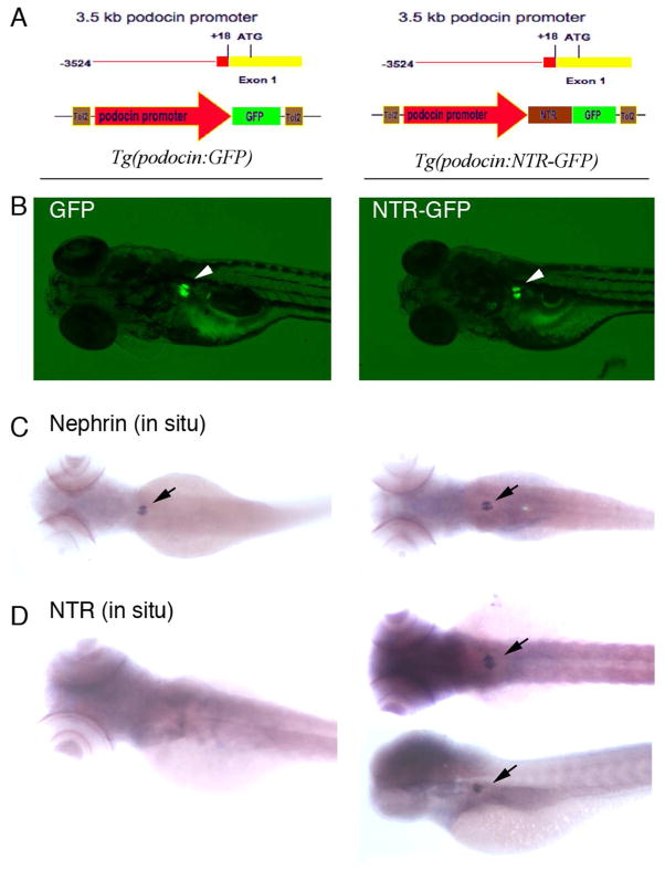
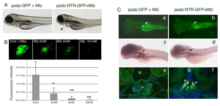
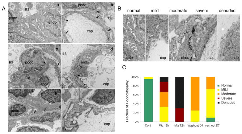
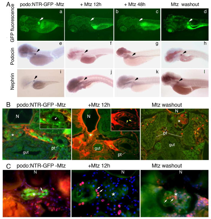
Podocytes are specialized cells that contribute critically to the normal structure and function of the glomerular filtration barrier. Their depletion plays an important role in the pathogenesis of glomerulosclerosis. Here, we report generation of a genetic model of conditional podocyte ablation and regeneration in zebrafish using a bacterial nitroreductase strategy to convert a prodrug, metronidazole, into a cytotoxic metabolite. A transgenic zebrafish line was generated that expresses green fluorescence protein (GFP) and the nitroreductase fusion protein under the control of the podocin promoter Tg(podocin:nitroreductase-GFP). Treatment of these transgenic zebrafish with metronidazole results in podocyte apoptosis, a loss of nephrin and podocin expression, foot process effacement, and a leaky glomerular filtration barrier. Following metronidazole washout, proliferating cells were detected in the glomeruli of recovering transgenic fish with a restoration of nitroreductase-GFP fluorescence, nephrin and podocin expression, a reestablishment of normal foot process architecture, and glomerular barrier function. Thus, our studies show that zebrafish podocytes are capable of regenerating following depletion, and establish the Tg(podocin:NTR-GFP) fish as a new model to study podocyte injury and repair.


Conflict of interest statement

COMPETING FINANCIAL INTERESTS

None

Figures

Fig. 1
Fig. 1. GFP and NTR-GFP expression under the 3.5-kb podocin promoter
A. Constructs used to generate the podocin driven GFP (left panel) and NTR-GFP (right panel) transgenic lines are shown respectively. B. GFP expression in glomeruli from transgenic fish Tg(podocin:GFP)(left panel, arrow) and Tg(podocin:NTR-GFP)(right panel, arrow). C. GFP expression (panels in B) overlaps with nephrin expression in glomeruli from Tg(podocin:GFP) (left panel, arrow) and Tg(podocin:NTR-GFP) (right panel, arrow) embryos. D. NTR is expressed in glomeruli from Tg(podocin:NTR-GFP)(right panel, arrows), but absent in glomeruli from Tg(podocin:GFP) embryos (left panel).
Fig. 2
Fig. 2. Metronidazole induces podocyte specific ablation
A. Mtz induced pericardial edema is seen in Tg(podocin:NTR-GFP) fish (right panel, indicated by the arrowhead) but not in Tg(podocin:GFP) fish (left panel). B. Treatment with Mtz results in attenuation and eventually loss of the GFP signal in the glomerulus of Tg(podocin:NTR-GFP) animals in a dose dependent manner (live images of glomeruli are shown in the upper panels and quantitation of fluorescence is shown in the lower graph). * indicates p<0.05, ** indicates p<0.01. C. A loss of GFP fluorescence and nephrin transcripts is seen in the glomeruli of Tg(podocin:NTR-GFP) fish (b and d respectively), but not in Tg(podocin:GFP)(a and c respectively) after treatment with Mtz for 12 hours. Arrows indicate the glomerulus. TUNEL-positive apoptotic cells (red) that co-label with the anti-panCrb antibody (green) are detected in the glomerulus of Mtz treated Tg(podocin:NTR-GFP) animals (f, arrows), but not in Mtz treated Tg(podocin:GFP) fish (e). N, notochord. * indicates the glomerulus.
Fig. 3
Fig. 3. Ultrastructural examination of podocytes in Mtz treated Tg(podocin:GFP) and Tg(podocin:NTR-GFP) fish larvae
A. Electron microscopy examination of Mtz treated Tg(podocin:GFP) larval fish shows normal podocyte morphology, intact foot processes and normal appearing glomerular basement membrane (a). Examination of Tg(podocin:NTR-GFP) fish treated for 12 hours with Mtz reveals the presence of foot process enfacement (b and c, indicated by arrows). Chromatin condensation and early nuclear fragmentation in podocytes is clearly seen at 12 hours post exposure to Mtz indicating podocyte apoptosis (c). A complete loss of foot process and significant podocyte destruction are observed in fish treated with Mtz for 72 hours (d), however, the morphology of neighboring endothelial cells appear grossly normal with intact intercellular junctional structures and glomerular basement membrane (b and d, arrowheads indicate intercellular junctional structures). 4 days after Mtz washout, foot process-like structures appear attached to the glomerular basement membrane in the glomerulus (arrows in e). 7 days after washout, near complete recovery of foot processes and slit diaphragms are found in the glomerulus (f). B. The change in podocyte ultrastructure in response to Mtz treatment and subsequent recovery after Mtz washout is categorized into mild, moderate, severe and denuded injuries based on established methods (25). C. Quantitation of damage of podocytes after Mtz treatment and podocyte recovery 4 day and 7 days after Mtz washout. Podo, podocyte. Endo, endothelial cell. BS, Bowman’s space. Cap, capillary space.
Fig. 4
Fig. 4. Dynamic changes in gene expression, glomerular function, and cell proliferation during Mtz induced podocyte injury and recovery after Mtz washout
A. GFP fluorescence is reduced and then absent after Mtz treatment for 12 and 48 hours, respectively (b–c), but reappears at 7 days after Mtz wash out (d). Arrows indicate the glomerulus. Whole mount in situ hybridization for podocin (f–h) and nephrin (j–l) transcripts reveals the concomitant loss and re-appearance of expression corresponding with the change in GFP fluorescence in the glomerulus of Mtz-treated Tg(podocin:NTR-GFP) animals. No change in GFP expression (a), or podocin (e) and nephrin (i) expression is seen in the glomerulus of Tg(podocin:NTR-GFP) animals without Mtz treatment. B. Assessment of glomerular filtration function using the rhodamine-conjugated albumin filtration assay. 5–6 hours after retro-orbital injection of rhodamine-albumin, rhodamine-albumin containing vesicles are detected inside pronephric tubule cells of Mtz treated Tg(Podocin:NTR-GFP) fish (middle panel, inset shows a higher magnified view of the tubule with arrowheads indicating red rhodamine-albumin containing vesicles inside the proximal tubular cells). Rhodamine-positive vesicles are not seen in untreated Tg(Podocin:NTR-GFP) control fish, although the uptake of freely filtered 10 kDa FITC-dextran is detected in these animals (left panel, arrowhead in inset indicates green FITC-dextran containing vesicles). No rhodamine-positive vesicles are seen in the pronephric tubule cells in recovered Tg(Podocin:NTR-GFP) animals at 7 days post Mtz washout (right panel). C. Detection of proliferating cells by BrdU incorporation. A small number of BrdU-positive cells are seen in the glomerulus of untreated Tg(podocin:NTR-GFP) laval fish (left panel, arrow indicates the red-colored BrdU signal in the nucleus. Green fluorescence marks NTR-GFP expressing podocytes). In Tg(Podocin:NTR-GFP) fish larvae treated with Mtz for 12 hours, despite the presence of BrdU labeling in neighboring cells of the glomerulus and pronephric tubules (arrows), almost no BrdU incorporation is detected in the glomerulus and the GFP fluorescence of podocytes is significant reduced (middle panel). Greatly increased BrdU staining is detected in the glomerulus of Tg(podocin:NTR-GFP) fish at 7 days post Mtz washout (right panel) (arrows). Overall GFP fluorescence is also increased in the glomerulus of these recovered animals. Some of the BrdU labeled cells are apparently also expressing GFP. N, notochord. Pt, pronephric tubule. * indicates glomerulus*.

References

    1. Asanuma K, Mundel P. The role of podocytes in glomerular pathobiology. Clin Exp Nephrol. 2003;7:255–259. - PubMed
    1. Fogo AB. Mechanisms of progression of chronic kidney disease. Pediatr Nephrol. 2007;22:2011–2022. - PMC - PubMed
    1. Moreno JA, Sanchez-Nino MD, Sanz AB, et al. A slit in podocyte death. Curr Med Chem. 2008;15:1645–1654. - PubMed
    1. Wiggins RC. The spectrum of podocytopathies: a unifying view of glomerular diseases. Kidney Int. 2007;71:1205–1214. - PubMed
    1. Hishiki T, Shirato I, Takahashi Y, et al. Podocyte injury predicts prognosis in patients with iga nephropathy using a small amount of renal biopsy tissue. Kidney Blood Press Res. 2001;24:99–104. - PubMed

Publication types

MeSH terms

LinkOut - more resources