. 2013 Mar 6;33(10):4241-52.
doi: 10.1523/JNEUROSCI.4893-12.2013.

Perineurial glia require Notch signaling during motor nerve development but not regeneration

Affiliations

Perineurial glia require Notch signaling during motor nerve development but not regeneration

Laura A Binari et al. J Neurosci. .

Abstract

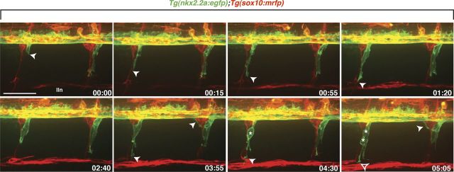
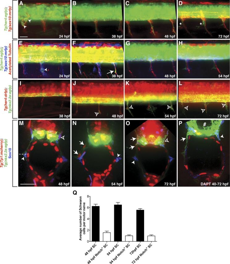
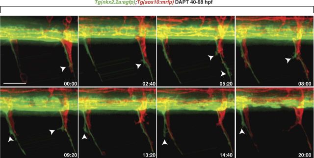
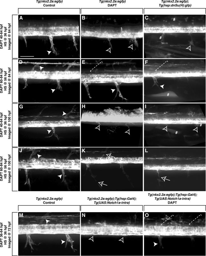
Motor nerves play the critical role of shunting information out of the CNS to targets in the periphery. Their formation requires the coordinated development of distinct cellular components, including motor axons and the Schwann cells and perineurial glia that ensheath them. During nervous system assembly, these glial cells must migrate long distances and terminally differentiate, ensuring the efficient propagation of action potentials. Although we know quite a bit about the mechanisms that control Schwann cell development during this process, nothing is known about the mechanisms that mediate the migration and differentiation of perineurial glia. Using in vivo imaging in zebrafish, we demonstrate that Notch signaling is required for both perineurial migration and differentiation during nerve formation, but not regeneration. Interestingly, loss of Notch signaling in perineurial cells also causes a failure of Schwann cell differentiation, demonstrating that Schwann cells require perineurial glia for aspects of their own development. These studies describe a novel mechanism that mediates multiple aspects of perineurial development and reveal the critical importance of perineurial glia for Schwann cell maturation and nerve formation.


Figures

Figure 1.
Figure 1.
Perineurial glia migrate as a chain out of the spinal cord. Frames captured from a 24 h time-lapse movie of a wild-type Tg(nkx2.2a:egfp);Tg(sox10:mrfp) larva beginning at 48 hpf. Numbers in lower right corners denote time elapsed from the first frame of the figure. At ∼50 hpf (00:00 time point), nkx2.2a+ perineurial glia (arrowhead) exited the spinal cord next to sox10+ (red) Schwann cells. Migrating perineurial cells had very few filopodia-like projections and traveled as a chain of cells along motor nerves. Once in the periphery, perineurial glial cell bodies often divided (asterisks) and lead cells had directed membrane processes (open arrowhead) while follower cells did not. Images are lateral views of the spinal cord with dorsal to the top and anterior to the left. lin, Lateral line nerve. Scale bar, 50 μm.
Figure 2.
Figure 2.
A–D, In Tg(sox10:mrfp);Tg(her4:egfp) larvae, some RFP+ Schwann cell precursors along peripheral motor nerves were GFP+ (arrowheads) at 24 hpf (A), but not between 38 and 72 hpf (B–D). D, At 72 hpf, Notch activity (asterisk) appeared in a pattern consistent with perineurial glial morphology. E–H, Notch activity was detected in Schwann cells and secondary motor axons (arrows) at 24 and 38 hpf in anti-acetylated tubulin labeled Tg(sox10:mrfp);Tg(her4:egfp) larvae, but not at 48 or 54 hpf. I–L, In Tg(nkx2.2a:egfp);Tg(her4:drfp) larvae, RFP+ perineurial glia (open arrowheads) populated motor nerves between 48 and 72 hpf. M–O, Transverse sections with dorsal to the top through the trunk of Tg(nkx2.2a:egfp);Tg(Tp1:mcherry) larvae labeled with an antibody specific to Sox10 (blue). At all stages, the majority of Schwann cells (arrowhead) were mCherry. mCherry+ Schwann cells (open arrowheads) were rarely seen. In contrast, at 48 and 72 hpf, all perineurial glia (arrows) were mCherry+. P, When Tg(nkx2.2a:egfp);Tg(Tp1:mcherry) larvae were treated with DAPT from 40 to 72 hpf, reporter line expression of mCherry was significantly reduced in the spinal cord (number sign). In these larvae, GFP+ perineurial glial processes (arrow) were observed as well as Sox10+ Schwann cells (arrowhead). Q, Quantification of Sox10+/ mCherry+ vs Sox10+/ mCherry Schwann cells (SC) along ventral motor roots to the horizontal myoseptum (horizontal dashed line). A–L, Lateral views of the spinal cord with dorsal to the top and anterior to the left. Scale bars: A–L, 50 μm; M–O, 25 μm.
Figure 3.
Figure 3.
Notch signaling is required for perineurial glial migration out of the spinal cord. Frames captured from a 20 h time-lapse movie of a DAPT-treated Tg(nkx2.2a:egfp);Tg(sox10:mrfp) larva beginning at 48 hpf. Numbers in lower right corners denote time elapsed from the first frame of the figure. At ∼50 hpf (00:00 time point), GFP+ perineurial processes (arrowhead) extended into the CNS at MEPs immediately next to Schwann cells (red). Over the time sequence, perineurial cell bodies never exited the spinal cord. Instead, their membrane protrusions got progressively more filamentous (arrowheads). Images are lateral views of the spinal cord with dorsal to the top and anterior to the left. Scale bar, 50 μm.
Figure 4.
Figure 4.
Notch is required during distinct stages of perineurial glial development. Lateral views of the spinal cord with dorsal to the top and anterior to the left of Tg(nkx2.2a:egfp) (A, B, D, E, G, H, J, K, M), Tg(nkx2.2a:egfp);Tg(hsp:dnSu(H):gfp) (C, F, I, L), and Tg(nkx2.2a:egfp);Tg(hsp:Gal4);Tg(UAS:Notch1a-intra) (N, O) larvae. A, At 64 hpf in control larvae, GFP+ perineurial cells (arrowhead) migrated into the PNS. In contrast, in larvae treated with DAPT from 40 to 64 hpf (B) or heat shocked at 36 hpf (C), perineurial cells failed to exit the CNS. Instead, only highly protrusive processes were detected (open arrowhead). D, By 84 hpf, GFP+ perineurial cells in control larvae populated both ventral and dorsal motor nerve braches. E, F, In contrast, in larvae treated with DAPT from 60 to 84 hpf (E) or heat shocked at 56 hpf (F), GFP+ perineurial cells only appeared to migrate along ventral motor nerves and were missing dorsally (dashed bracket). G, In 100 hpf control larvae, GFP+ perineurial cells were found on dorsal and ventral motor nerves. H, I, In larvae treated with DAPT from 40 to 64 hpf (H) or heat shocked at 36 hpf (I) and allowed to recover until 100 hpf, perineurial processes but not cell bodies were seen in the periphery. K, L, In contrast, in larvae treated with DAPT from 60 to 84 hpf (K) or heat shocked at 56 hpf (L) and allowed to recover until 100 hpf, GFP+ perineurial cells were associated with peripheral motor nerves in discontinuous chains (arrows), which was never observed in controls (J) (arrowheads). M, N, When compared with control larvae at 72 hpf (M), heat shock induction of a constitutively active form of the Notch intracellular domain at 36 hpf (N) resulted in a failure of perineurial glial migration into the periphery. O, When treated with DAPT and heat shocked at 36 hpf, partial rescue of the perineurial glial migration phenotype was observed at 72 hpf. Number sign denotes examples of GFP+ cells due to heat shock. Scale bar, 50 μm.
Figure 5.
Figure 5.
Notch signaling is required for perineurial glial differentiation. A, C, E, Lateral views of the spinal cord with dorsal to the top and anterior to the left of Tg(nkx2.2a:egfp);Tg(olig2:dsred) larvae at 6 dpf. B, D, F, Transverse sections with dorsal to the top through the trunk of 6 dpf Tg(nkx2.2a:egfp) larvae labeled with an antibody specific to ZO-1 (red). A, In control larvae, GFP+ perineurial cells (arrows) ensheathed dorsal and ventral motor nerves. B, Within these perineurial cells, high levels of ZO-1+ tight junctions (arrowhead) were detected extending from the ventral spinal cord to the horizontal myoseptum (dashed line). C, D, In contrast, when larvae were treated with DAPT from 40 to 64 hpf and left to recover until 6 dpf, perineurial glial processes but not cell bodies migrated into the periphery and no ZO-1+ tight junctions were detected. E, In larvae treated from 60 to 84 hpf and allowed to recover until 6 dpf, GFP+ perineurial cells migrated along motor nerves, but not in continuous chains (arrow). F, In this instance, ZO-1+ tight junctions were observed in perineurial cells most proximal to the ventral spinal cord, but never further along the nerve. B, D, F, Insets show magnified view of the dashed rectangular regions. Scale bars: A, C, E, 50 μm; B, D, F, 25 μm.
Figure 6.
Figure 6.
Perturbed perineurial glial development influences Schwann cell development. A–D, Transverse sections with dorsal to the top through the trunk of Tg(nkx2.2a:egfp);Tg(olig2:dsred) larvae labeled with an antibody specific to Sox10 (blue). F–K, Transverse (F–H) and whole-mount (I–K) views with dorsal to the top of mbp expression in 96 hpf larvae. A–D, Compared with controls, DAPT-treated larvae had significantly fewer Schwann cells (arrowheads) populating the motor nerve. E, Quantification of Sox10+ cells along ventral motor roots to the horizontal myoseptum. F, I, In control larvae, mbp transcript was detected in oligodendrocytes in the CNS (arrows) as well as in Schwann cells along motor nerves (open arrowheads) and the PLLn (arrowheads). G, J, In contrast, larvae treated with DAPT from 40 to 72 hpf showed strongly reduced mbp expression in the CNS and no detectable expression along motor nerves. However, PLLn expression was indistinguishable from controls. H, K, Similar to the early DAPT treatment, larvae exposed to DAPT from 60 to 72 hpf showed no motor nerve mbp expression, but indistinguishable expression in the CNS and along the PLLn. Statistical significance was determined using the unpaired t test. p-values are shown for each treatment type compared with controls. Horizontal dashed line denotes horizontal myoseptum. Scale bars: A–D, 25 μm; F–H, 25 μm; I–K, 50 μm.
Figure 7.
Figure 7.
Notch activity is not detected in perineurial glia after injury. A, B, Images taken from a Tg(nkx2.2a:egfp);Tg(her4:drfp) (A) and Tg(nkx2.2a:egfp);Tg(Tp1:mcherry) (B) larva after motor nerve injury beginning at 6 dpf. Numbers in lower right corners denote time elapsed from the first frame of the figure. A, B, Motor nerve-associated perineurial glia (asterisks) never express RFP or mCherry after nerve injury. A, Number sign denotes RFP expression in the spinal cord. B, 1–4 mark mCherry+ cells in the periphery. Brackets show injury zones. All images are lateral views of a motor nerve with dorsal to the top and anterior to the left. Scale bar, 10 μm.

References

    1. Akert K, Sandri C, Weibel ER, Peper K, Moor H. The fine structure of the perineural endothelium. Cell Tissue Res. 1976;165:281–295. - PubMed
    1. Andersson ER, Sandberg R, Lendahl U. Notch signaling: simplicity in design, versatility in function. Development. 2011;138:3593–3612. - PubMed
    1. Banerjee S, Gordon L, Donn TM, Berti C, Moens CB, Burden SJ, Granato M. A novel role for MuSK and non-canonical Wnt signaling during segmental neural crest cell migration. Development. 2011;138:3287–3296. - PMC - PubMed
    1. Bourne GH. The structure and function of nervous tissue. New York: Academic; 1968.
    1. Bray SJ. Notch signalling: a simple pathway becomes complex. Nat Rev Mol Cell Biol. 2006;7:678–689. - PubMed

Publication types

MeSH terms

Substances

LinkOut - more resources