. 2013 Apr 26;288(17):11751-60.
doi: 10.1074/jbc.M112.434365. Epub 2013 Mar 6.

Minor modifications to the phosphate groups and the C3' acyl chain length of lipid A in two Bordetella pertussis strains, BP338 and 18-323, independently affect Toll-like receptor 4 protein activation

Affiliations

Minor modifications to the phosphate groups and the C3' acyl chain length of lipid A in two Bordetella pertussis strains, BP338 and 18-323, independently affect Toll-like receptor 4 protein activation

Nita R Shah et al. J Biol Chem. .

Abstract

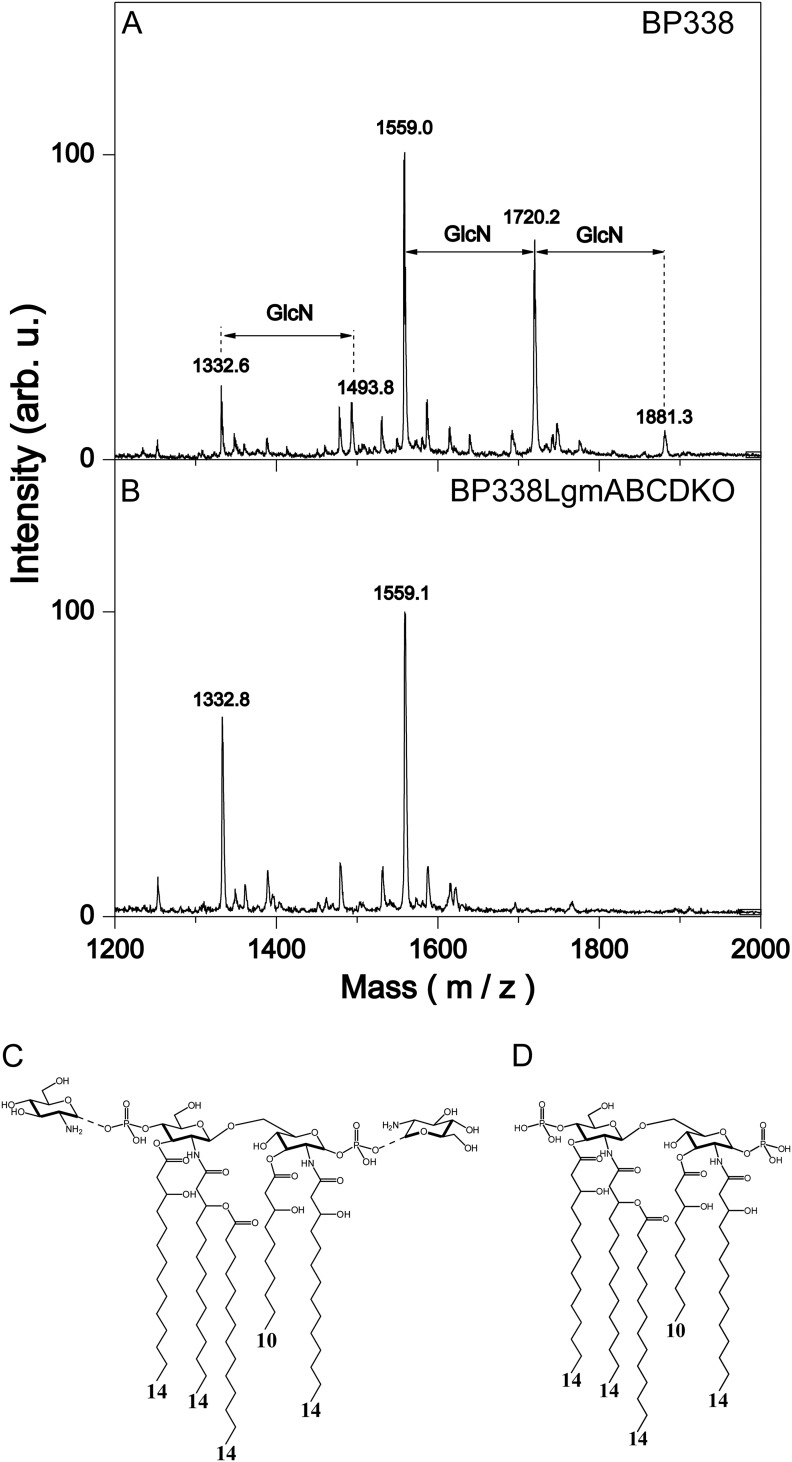
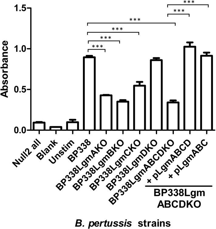
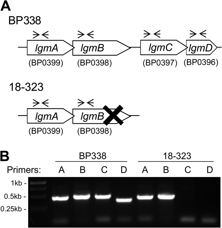
Lipopolysaccharides (LPS) of Bordetella pertussis are important modulators of the immune system. Interaction of the lipid A region of LPS with the Toll-like receptor 4 (TLR4) complex causes dimerization of TLR4 and activation of downstream nuclear factor κB (NFκB), which can lead to inflammation. We have previously shown that two strains of B. pertussis, BP338 (a Tohama I-derivative) and 18-323, display two differences in lipid A structure. 1) BP338 can modify the 1- and 4'-phosphates by the addition of glucosamine (GlcN), whereas 18-323 cannot, and 2) the C3' acyl chain in BP338 is 14 carbons long, but only 10 or 12 carbons long in 18-323. In addition, BP338 lipid A can activate TLR4 to a greater extent than 18-323 lipid A. Here we set out to determine the genetic reasons for the differences in these lipid A structures and the contribution of each structural difference to the ability of lipid A to activate TLR4. We show that three genes of the lipid A GlcN modification (Lgm) locus, lgmA, lgmB, and lgmC (previously locus tags BP0399-BP0397), are required for GlcN modification and a single amino acid difference in LpxA is responsible for the difference in C3' acyl chain length. Furthermore, by introducing lipid A-modifying genes into 18-323 to generate isogenic strains with varying penta-acyl lipid A structures, we determined that both modifications increase TLR4 activation, although the GlcN modification plays a dominant role. These results shed light on how TLR4 may interact with penta-acyl lipid A species.


Figures

FIGURE 1.
FIGURE 1.
Structural analysis of lipid A with negative-ion MALDI mass spectrum analysis. Mass spectra of BP338 (A) and BP338LgmABCDKO (B), the full lgm locus deletion mutant. Peaks at m/z 1559 represent penta-acyl lipid A that lack GlcN modification, peaks at m/z 1720 represent penta-acyl lipid A with one GlcN modification at either phosphate group, and peaks at m/z 1881 represent penta-acyl lipid A with a GlcN modification at both phosphate groups. The peaks at m/z 1333 and 1494 represent tetra-acyl species. arb. u., arbitrary units. C and D, the lipid A structures present in BP338 (C) and BP338LgmABCDKO (D) as derived by mass spectral analysis. The numbers at the bottom of the structures indicate the length of the acyl chains.
FIGURE 2.
FIGURE 2.
TLR4 activity of heat-killed bacterial cells as measured with the HEK-Blue NFκB TLR4 activity assay. Null2 all, stimulation of HEK-Blue Null2 cell line that lacks TLR4 expression with all LPS variants; Blank, medium only with no HEK-Blue cells; Unstim, HEK-Blue hTLR4 cells stimulated with medium only; BP338LgmABCDKO, the full Lgm locus deletion mutant; BP338LgmABCDKO + pLgmABCD, full Lgm locus deletion mutant complemented with lgmABCD; BP338LgmABCDKO + pLgmABC, full Lgm locus deletion mutant complemented with lgmABC. Graph shows the results of one representative experiment of three, n = 6 replicates per experiment. p values: < 0.001 (***). Error bars indicate S.D.
FIGURE 3.
FIGURE 3.
Genetic analysis of the Lgm locus BP338 and 18-323 strains of B. pertussis. A, the Lgm loci of BP338 and 18-323, as determined by sequence analysis. These data were provided by the pathogen genomics group at the Wellcome Trust Sanger Institute and can be obtained from the Sanger Institute website. X represents a TT deletion mutation at bp 981 of lgmB in 18-323; the forward arrows and the reverse arrows illustrate the annealing sites of the primers used for the PCR shown in B. B, PCR of the Lgm locus genes in BP338 and 18-323 using gene-specific primers. Expected positive bands: 0.48 kb (lgmA primers), 0.51 kb (lgmB primers), 0.50 kb (lgmC primers), and 0.40 kb (lgmD primers).
FIGURE 4.
FIGURE 4.
Negative-ion MALDI mass spectra and structure of lipid A from B. pertussis 18-323 strains containing BP338 lipid A-modifying genes. A–D, 18-323 (A), 18-323 + pPtacLgmABCD (complemented with the Lgm locus of BP338) (B), 18-323 + pPtacLpxA338 (complemented with lpxA of BP338) (C), and 18-323 + pPtacLgmABCDLpxA338 (complemented with the Lgm locus of BP338 and lpxA of BP338) (D). Arrows labeled with C10OH, C12OH, or C14OH indicate the 10-, 12-, or 14-carbon acyl chains absent in the tetra-acyl and present in the respective penta-acyl lipid A species. Arrows labeled with GlcN indicate the addition of GlcN at a phosphate group. The lipid A structures are summarized to the right of the mass spectra. Numbers at the bottom of the structures indicate the length of the acyl chains. Structures with GlcN modifications (B and D) have one GlcN added to either of the phosphate groups (peaks at m/z 1664, 1692, or 1720). arb. u., arbitrary units.
FIGURE 5.
FIGURE 5.
Alignment of LpxA sequences from various Gram-negative species. Species (top to bottom) are: E. coli K-12 (EC), P. aeruginosa (PA), B. bronchiseptica (Bbronch), Bordetella parapertussis (Bpara), B. pertussis Tohama I BP338 (Tohama), B. pertussis 18-323 (18323), R. sphaeroides (RS), and P. gingivalis (Pging). The alignment was constructed with ClustalW (32). The down arrow indicates the single amino acid difference between LpxA of BP338 and 18-323 (amino acid 173), which corresponds to Gly-176 in E. coli LpxA. The bottom right inset shows the active site region between chains A (gray) and B (brown) of the E. coli LpxA homo-trimer and the amino acids surrounding the 14-carbon acyl chain (pink). E. coli LpxA amino acids: His-160 (green), Gly-173 (blue), Gly-176 (red), and His-191 (orange). E. coli LpxA structure PDB ID 2QIA (9) was used to generate this figure in Swiss-Pdb Viewer (ExPASy) (33).
FIGURE 6.
FIGURE 6.
TLR4 activity of purified LPS from 18-323 strains as measured with the HEK-Blue NFκB TLR4 activity assay. Null2 all, stimulation of HEK-Blue Null2 cell line that lacks TLR4 expression with all LPS variants; Blank, medium only with no HEK-Blue cells; Unstim, HEK-Blue hTLR4 cells stimulated with medium only; 18-323 + pPtacLpxA338, 18-323 complemented with lpxA from BP338; 18-323 + pPtacLgmABCD, 18-323 complemented with Lgm locus from BP338; 18-323 + pPtacLgmABCDLpxA338, 18-323 complemented with both lpxA and Lgm locus from BP338. Graph shows the results of one representative experiment of three, n = 6. p values: <0.01 (**), < 0.001 (***). Error bars indicate S.D.

References

    1. Trent M. S., Stead C. M., Tran A. X., Hankins J. V. (2006) Diversity of endotoxin and its impact on pathogenesis. J. Endotoxin Res. 12, 205–223 - PubMed
    1. Caroff M., Brisson J., Martin A., Karibian D. (2000) Structure of the Bordetella pertussis 1414 endotoxin. FEBS Lett. 477, 8–14 - PubMed
    1. Caroff M., Karibian D. (2003) Structure of bacterial lipopolysaccharides. Carbohydr. Res. 338, 2431–2447 - PubMed
    1. Raetz C. R., Reynolds C. M., Trent M. S., Bishop R. E. (2007) Lipid A modification systems in Gram-negative bacteria. Annu. Rev. Biochem. 76, 295–329 - PMC - PubMed
    1. Trent M. S., Ribeiro A. A., Lin S., Cotter R. J., Raetz C. R. (2001) An inner membrane enzyme in Salmonella and Escherichia coli that transfers 4-amino-4-deoxy-l-arabinose to lipid A: induction on polymyxin-resistant mutants and role of a novel lipid-linked donor. J. Biol. Chem. 276, 43122–43131 - PubMed

Publication types