

Accelerated ischemic vascular retinopathy after intravitreally injected bevacizumab for central retinal vein occlusion in elderly patients
- PMID: 23467497
- PMCID: PMC3589185
- DOI: 10.2147/OPTH.S30156
Accelerated ischemic vascular retinopathy after intravitreally injected bevacizumab for central retinal vein occlusion in elderly patients
Abstract
Background: Ischemic changes in the retinal circulation are an uncommon but severe adverse vascular reaction to intravitreal bevacizumab (Avastin(®), Genentech, San Francisco, CA, USA/Roche, Basel, Switzerland) for central retinal vein occlusion (CRVO). In the two cases reported here, ischemic changes in the retina vasculature following intravitreal bevacizumab for CRVO were observed with the aim of describing the clinical and angiographic features of these changes.
Methods: Two elderly patients with recent-onset CRVO received one off-label intravitreal injection of bevacizumab 0.05 mL/1.25 mg.
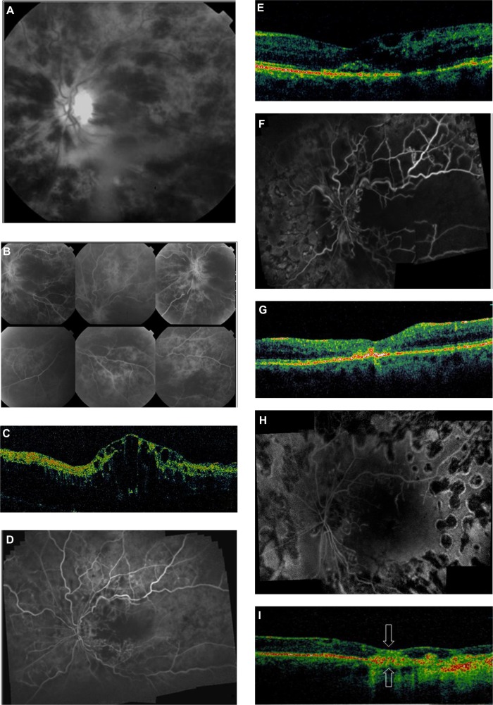
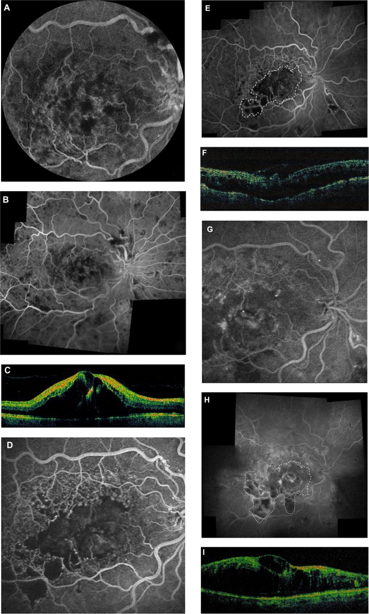
Results: In Case 1, the patient's pre-treatment visual acuity was 20/400. At 3 weeks post injection, the patient could count fingers at a distance of 1 ft (30 cm) and fluorescein angiography showed reduction in intraretinal hemorrhages and areas of retinal non-perfusion. However, at 6 weeks these were markedly increased compared with those seen in the photograph taken 3 weeks after treatment. In Case 2, the patient's pre-treatment visual acuity was 20/200. At 1 month post injection, vision had decreased to 20/400 and fluorescein angiography showed severe macular ischemia with a remarkable capillary dropout throughout the macula.
Conclusion: Ischemic retinal injury may be an uncommon but severe adverse vascular reaction to intravitreal bevacizumab for CRVO. Although progression of retinal ischemia in CRVO could be observed shortly after intravitreal bevacizumab, whether this is a drug- or procedure-related effect or part of the natural history of the condition remains uncertain.
Keywords: Avastin; intraretinal hemorrhage; ischemia; macular infarction; retinal non-perfusion.
Figures
Similar articles
-
Prospective study of intravitreal ranibizumab as a treatment for decreased visual acuity secondary to central retinal vein occlusion.Am J Ophthalmol. 2009 Feb;147(2):298-306. doi: 10.1016/j.ajo.2008.08.016. Epub 2008 Oct 17. Am J Ophthalmol. 2009. PMID: 18929354 Clinical Trial.
-
Intravitreal bevacizumab (Avastin) treatment of macular edema in central retinal vein occlusion: a short-term study.Retina. 2006 Mar;26(3):279-84. doi: 10.1097/00006982-200603000-00005. Retina. 2006. PMID: 16508427
-
Aflibercept for Previously Treated Macular Edema Associated with Central Retinal Vein Occlusions: 1-Year Results of the NEWTON Study.Ophthalmol Retina. 2018 Feb;2(2):128-133. doi: 10.1016/j.oret.2017.05.017. Epub 2017 Oct 12. Ophthalmol Retina. 2018. PMID: 31047339 Clinical Trial.
-
Long-term effect of early intervention with single intravitreal injection of bevacizumab followed by panretinal and macular grid photocoagulation in central retinal vein occlusion (CRVO) with macular edema: a pilot study.Eye (Lond). 2011 Feb;25(2):239-44. doi: 10.1038/eye.2010.225. Eye (Lond). 2011. PMID: 21307880 Free PMC article.
-
Intravitreal bevacizumab injections for treatment of central retinal vein occlusion: six-month results of a prospective trial.Retina. 2007 Oct;27(8):1004-12. doi: 10.1097/IAE.0b013e3180ed458d. Retina. 2007. PMID: 18040236 Clinical Trial.
Cited by
-
A pharmacological approach in newly established retinal vein occlusion model.Sci Rep. 2017 Mar 2;7:43509. doi: 10.1038/srep43509. Sci Rep. 2017. PMID: 28252108 Free PMC article.
-
Acute macular neuroretinopathy associated with intravitreal anti-VEGF injection: A case report.Am J Ophthalmol Case Rep. 2022 Aug 18;28:101687. doi: 10.1016/j.ajoc.2022.101687. eCollection 2022 Dec. Am J Ophthalmol Case Rep. 2022. PMID: 36046518 Free PMC article.
-
Analysis of Potential Ischemic Effect of Intravitreal Bevacizumab on Unaffected Retina in Treatment-Naïve Macular Edema Due to Branch Retinal Vein Occlusion: A Prospective, Interventional Case-Series.PLoS One. 2016 Sep 12;11(9):e0162533. doi: 10.1371/journal.pone.0162533. eCollection 2016. PLoS One. 2016. PMID: 27618696 Free PMC article.
-
Hemorrhagic macular infarction after intravitreal bevacizumab for chronic multifocal central serous chorioretinopathy.Case Rep Ophthalmol. 2014 Jul 11;5(2):207-11. doi: 10.1159/000365442. eCollection 2014 May. Case Rep Ophthalmol. 2014. PMID: 25126075 Free PMC article.
References
-
- Iturralde D, Spaide RF, Meyerle CB, et al. Intravitreal bevacizumab (Avastin) treatment of macular edema in central retinal vein occlusion: a short-term study. Retina. 2006;26(3):279–284. - PubMed
-
- Kriechbaum K, Michels S, Prager F, et al. Intravitreal Avastin for macular oedema secondary to retinal vein occlusion: a prospective study. Br J Ophthalmol. 2008;92(4):518–522. - PubMed
-
- Peters S, Heiduschka P, Julien S, et al. Ultrastructural findings in the primate eye after intravitreal injection of bevacizumab. Am J Ophthalmol. 2007;143(6):995–1002. - PubMed
-
- Lee CS, Koh HJ. Multiple retinal haemorrhages in diabetic retinopathy after adjunctive intravitreal bevacizumab (Avastin) with pars plana vitrectomy. Acta Ophthalmol. 2008;86(7):812–813. - PubMed
-
- Hayreh SS, Zimmerman MB, Podhajsky P. Incidence of various types of retinal vein occlusion and their recurrence and demographic characteristics. Am J Ophthalmol. 1994;117(4):429–441. - PubMed
Publication types
LinkOut - more resources
Full Text Sources
Other Literature Sources
