. 2013 Mar 15;73(6):1800-10.
doi: 10.1158/0008-5472.CAN-12-1916. Epub 2013 Mar 6.

Arkadia regulates tumor metastasis by modulation of the TGF-β pathway

Affiliations

Arkadia regulates tumor metastasis by modulation of the TGF-β pathway

Marco A Briones-Orta et al. Cancer Res. .

Abstract

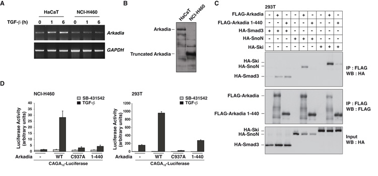
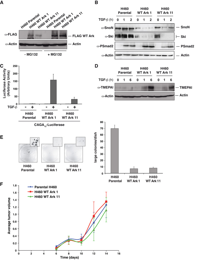
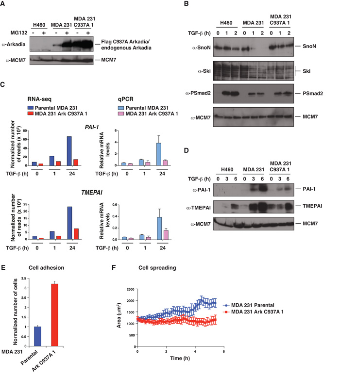
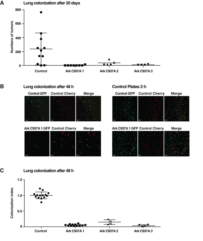
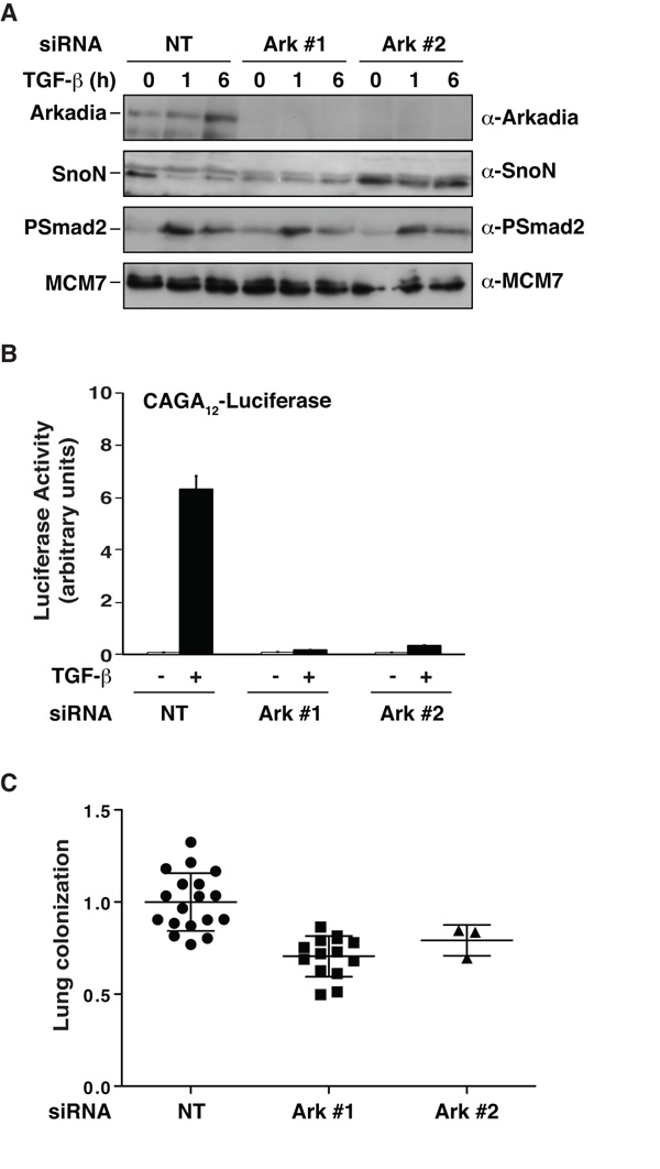
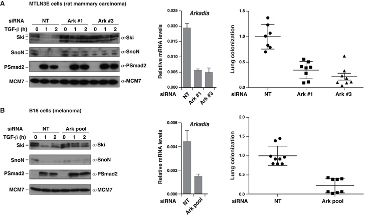
TGF-β can act as a tumor suppressor at early stages of cancer progression and as a tumor promoter at later stages. The E3 ubiquitin ligase Arkadia (RNF111) is a critical component of the TGF-β signaling pathway, being required for a subset of responses, those mediated by Smad3-Smad4 complexes. It acts by mediating ligand-induced degradation of Ski and SnoN (SKIL), which are 2 potent transcriptional repressors. Here, we investigate the role of Arkadia in cancer using model systems to address both potential tumor-suppressive and tumor-promoting roles. Stable reexpression of Arkadia in lung carcinoma NCI-H460 cells, which we show contain a hemizygous nonsense mutation in the Arkadia/RNF111 gene, efficiently restored TGF-β-induced Smad3-dependent transcription, and substantially decreased the ability of these cells to grow in soft agar in vitro. However, it had no effect on tumor growth in vivo in mouse models. Moreover, loss of Arkadia in cancer cell lines and human tumors is rare, arguing against a prominent tumor-suppressive role. In contrast, we have uncovered a potent tumor-promoting function for Arkadia. Using 3 different cancer cell lines whose tumorigenic properties are driven by TGF-β signaling, we show that loss of Arkadia function, either by overexpression of dominant negative Arkadia or by siRNA-induced knockdown, substantially inhibited lung colonization in tail vein injection experiments in immunodeficient mice. Our findings indicate that Arkadia is not critical for regulating tumor growth per se, but is required for the early stages of cancer cell colonization at the sites of metastasis.


Figures

Figure 1
Figure 1. NCI-H460 cells express a truncated and catalytically inactive version of Arkadia
A. Levels of Arkadia and GAPDH mRNA in HaCaT and NCI-H460 cells were assayed by RT-PCR. B. Whole cell extracts from HaCaT and NCI-H460 cells analyzed by Western Blot using an antibody against Arkadia. C. Whole cell extracts from 293T cells transfected with the plasmids indicated were immunoprecipitated (IP) using anti-FLAG beads and Western blotted (WB) using HA and FLAG antibodies. Inputs show expression levels of Arkadia, Smad3, Ski and SnoN. D. NCI-H460 and 293T cells were transfected with the CAGA12-Luciferease reporter and the TK-Renilla control along with plasmids expressing wild-type (WT), C937A, or truncated (1–440) Arkadia. Luciferase activity was normalized to Renilla activity. Means and standard deviations are shown of representative experiments performed in triplicate.
Figure 2
Figure 2. Reintroduction of Arkadia in NCI-H460 cells restores TGF-β-dependent Ski/SnoN degradation, transcriptional responses and inhibits anchorage independent growth, but has no effect on their tumorigenic properties in vivo
A. Western blot showing Arkadia levels in two NCI-H460-derived cell lines expressing FLAG-tagged Arkadia. Cells were treated for 4 h ± MG132. Actin is a loading control. B. Whole cell extracts from cells treated ± TGF-β Western blotted using the antibodies indicated. C. Luciferase assays were performed as in Fig. 1. D. Whole cell extracts prepared from the cell lines indicated and Western blotted using antibodies shown. E. NCI-H460 parental cells or the clones expressing FLAG-tagged Arkadia were grown in soft agar for 10 days. The numbers of colonies were quantified and are shown in the bar graph (right). F. The cell lines shown were injected subcutaneously in both flanks of Balbc nu/nu mice. The data are the averages of the tumor volumes for four mice (8 tumors in total) per cell line.
Figure 3
Figure 3. Expression of Arkadia C937A abolishes Arkadia function in MDA-MB-231 cells and alters their adhesion and spreading properties
A–D. NCI-H460 cells, parental MDA-MB-231 cells, and a stable MDA-MB-231 cell line expressing FLAG-tagged Arkadia C937A (clone 1) were treated ± MG132 for 4 h (A) or with TGF-β for the times indicated (B–D). Whole cell extracts were Western blotted using the antibodies indicated (A, B and D). In C, levels of mRNA for the genes shown were analyzed either by RNA-seq or by qPCR which was normalized to GAPDH. E. GFP-labeled parental MDA-MB-231 cells and Arkadia C937A cells (clone 1) were mixed with an equal number of parental cells expressing mCherry, seeded on a monolayer of HUVEC cells and incubated for 1 h, after which they were washed, fixed and counted. The mean ratio of GFP:mCherry cells and standard deviations from 6 wells is plotted. F. GFP-labeled parental MDA-MB-231 cells or Arkadia C937A cells (clone 1) were seeded on a layer of HUVEC cells, and movies were recorded. Cell spreading was measured by quantification of changes in the area of the cells over time using the Imaris software. The graphs show the means of the average cell area for 4 independent wells for each cell type with standard errors. Similar results were obtained in three independent experiments.
Figure 4
Figure 4. Inhibition of Arkadia activity in MDA-MB-231 by expression of Arkadia C937A inhibits lung colonization
A. Parental MDA-MB-231 and three independent clones of Arkadia C937A-expressing cells were injected into the tail vein of immunodeficient mice. After 20 days, lungs were analyzed for number of tumors. B and C. GFP-labeled parental MDA-MB-231 cells and the three independent clones of Arkadia C937A-expressing cells were mixed with an equal number of parental cells expressing mCherry and injected into the tail vein of immunodeficient mice. After 2 days mice were culled and lungs directly analyzed by confocal microscopy. Representative pictures of tumor cell distribution for parental and one clone of Arkadia C937A-expressing cells were taken (B, left panels). As a control, aliquots of the mixed GFP/mCherry cells were plated prior to injection (B, right panels). The magnification scale bar corresponds to 100 μm. The ratio between the GFP/mCherry cells in the lungs from multiple mouse experiments is plotted (C). In A and C each point represents data from a single mouse, and means and standard deviation are plotted.
Figure 5
Figure 5. The ability of MDA-MB-231 cells to colonize lungs requires Arkadia function
A–C. GFP and mCherry-labeled MDA-MB-231 cells were transfected with a control (NT), and GFP-labeled MDA-MB-231 cells were transfected with two different siRNAs against Arkadia (Ark #1 and #2). 24 h later cells were split for immunoblots, CAGA12-Luciferase reporter assays and mice experiments. A. Whole cell extracts were prepared from the GFP-labeled cells treated ± TGF-β and Western blotted using the antibodies indicated. B. The GFP-labeled cells were transfected with the CAGA12-Luciferase and TK-Renilla reporters then treated ± TGF-β. Coupled Luciferase/Renilla assays were as in Fig. 1. C. Equal numbers of NT mCherry cells were mixed with either NT GFP cells or Ark #1 and Ark #2 siRNA GFP cells and were injected into the tail vein of immunodeficient mice. Analysis and quantitation of the experiment was as in Fig. 4.
Figure 6
Figure 6. MTLN3E and B16 cells require Arkadia to colonize lungs
A and B. GFP and mCherry-labeled MTLN3E cells or B16 cells were transfected with a control (NT), and GFP-labeled MTLN3E and B16 cells were transfected with siRNAs against Arkadia as indicated. 24 h later cells were split for immunoblots, qPCR for Arkadia/GAPDH and 48-h lung colonization mouse experiments, which were as described in Fig. 4.

References

    1. Hanahan D, Weinberg RA. Hallmarks of cancer: the next generation. Cell. 2011;144:646–74. - PubMed
    1. Massague J. TGFβ in Cancer. Cell. 2008;134:215–30. - PMC - PubMed
    1. Padua D, Massague J. Roles of TGFβ in metastasis. Cell Res. 2009;19:89–102. - PubMed
    1. Gupta GP, Massague J. Cancer metastasis: building a framework. Cell. 2006;127:679–95. - PubMed
    1. Feng XH, Derynck R. Specificity and versatility in TGF-β signaling through Smads. Annu Rev Cell Dev Biol. 2005;21:659–93. - PubMed

Publication types