. 2013 Jun;378(1-2):153-9.
doi: 10.1007/s11010-013-1606-x. Epub 2013 Mar 7.

MicroRNA-1 prevents high-fat diet-induced endothelial permeability in apoE knock-out mice

Affiliations

MicroRNA-1 prevents high-fat diet-induced endothelial permeability in apoE knock-out mice

Hua Wang et al. Mol Cell Biochem. 2013 Jun.

Abstract

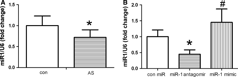
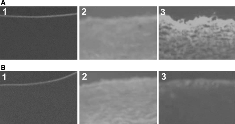
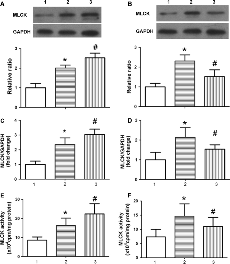
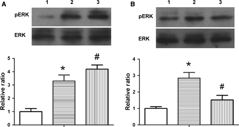
The development of atherosclerosis (AS) is a multifactorial process in which elevated plasma cholesterol levels play a central role. As a new class of players involved in AS, the regulation and function of microRNAs (miR) in response to AS remain poorly understood. This study analyzed the effects of miR-1 (antagomir and mimic) on endothelial permeability and myosin light chain kinase (MLCK) expression and activity in the artery wall of apoE knock-out mice after feeding them a high-cholesterol diet. Further, we tested to determine whether that effects are involved in ERK phosphorylation. Here, we show that a high-cholesterol diet induces a significant decrease of miR-1 expression. Histopathologic examination demonstrated that miR-1 antagomir enhances endothelial permeability induced by high cholesterol and miR-1 mimic attenuated endothelial barrier dysfunction. Consistent with endothelial permeability, Western blotting, qPCR, and γ-(32)P-ATP phosphate incorporation showed that MLCK expression and activity were further increased in miR-1 antagomir-treated mice and decreased in miR-1 mimic-treated mice compared with those of mice receiving control miR. Further mechanistic studies showed that high-cholesterol-induced extracellular signal regulated kinase (ERK) activation was enhanced by miR-1 antagomir and attenuated by miR-1 mimic. Collectively, those results indicate that miR-1 contributes to endothelial barrier function via mechanisms involving not only MLCK expression and activity but also ERK phosphorylation.


Figures

Fig. 1
Fig. 1
Total RNA was extracted from the aorta of different groups using TRIzol reagent; MiR-1 expression was determined using the miRNA plate assay kit; for normalized RNA content, the U6 snRNA was the internal control. a miR-1 expression was detected in control and AS mice. b miR-1 treatment influences miR-1 expression in the aorta. Levels of miR-1 were detected in different miR-treated AS mice, *P < 0.05 significance relative to Con, # P < 0.05 significance relative to miR-1 antagomir
Fig. 2
Fig. 2
miR-1 treatment influences endothelial permeability in the aorta. After aorta intimas were incubated with NHSLC-biotin for 30 min, frozen sections were incubated with XRITC-avidin to localize surface-bound biotin. Photographs taken at 20-fold magnification. a 1: Con + Con miR; 2: AS + Con miR; 3: AS + miR-1 antagomir; B: 1: Con + Con miR; 2: AS + Con miR; 3: AS + miR-1 mimic; *P < 0.05 significance relative to Con, # P < 0.05 significance relative to AS + Con miR
Fig. 3
Fig. 3
miR-1 treatment influences MLCK expression and activity in the aorta. Western blotting (a, b) and qPCR (c, d) were performed as described in the “Materials and methods” section to detect MLCK expression, and γ-32P-ATP phosphate incorporation was performed to assay MLCK activity(E and F). A,C,E: 1:Con + Con miR; 2: AS + Con miR; 3: AS + miR-1 antagomir; B,D,F: 1:Con + Con miR; 2: AS + Con miR; 3: AS + miR-1 mimic; *P < 0.05 significance relative to Con, # P < 0.05 significance relative to AS + Con miR
Fig. 4
Fig. 4
miR-1 treatment influences ERK phosphorylation in the aorta. Control and AS mice were injected with different miRs. Four weeks later, phosphorylated ERK (pERK) and total ERK (ERK) were determined in aortic tissues by western blot analysis. The upper panel is a representative western blot for pERK and ERK and the lower panel is the quantitative data from western blot analysis for the ratio of pERK/ERK. a 1: Con + Con miR; 2: AS + Con miR; 3: AS + miR-1 antagomir; b 1: Con + Con miR; 2: AS + Con miR; 3: AS + miR-1 mimic; *P < 0.05 significance relative to Con, # P < 0.05 significance relative to AS + Con miR

References

    1. Thum T. MicroRNA therapeutics in cardiovascular medicine. EMBO Mol Med. 2012;4:3–14. doi: 10.1002/emmm.201100191. - DOI - PMC - PubMed
    1. Libby P, Ridker PM, Hansson GK. Progress and challenges in translating the biology of atherosclerosis. Nature. 2011;473:317–325. doi: 10.1038/nature10146. - DOI - PubMed
    1. Collins NT, Cummins PM, Colgan OC, et al. Cyclic strain-mediated regulation of vascular endothelial occludin and ZO-1: influence on intercellular tight junction assembly and function. Arterioscler Thromb Vasc Biol. 2006;26:62–68. doi: 10.1161/01.ATV.0000194097.92824.b3. - DOI - PubMed
    1. Huang Q, Xu W, Ustinova E, et al. Myosin light chain kinase-dependent microvascular hyperpermeability in thermal injury. Shock. 2003;20:363–368. doi: 10.1097/01.shk.0000079425.0000.db. - DOI - PubMed
    1. Stroka KM, Aranda-Espinoza H. Endothelial cell substrate stiffness influences neutrophil transmigration via myosin light chain kinase-dependent cell contraction. Blood. 2011;118:1632–1640. doi: 10.1182/blood-2010-11-321125. - DOI - PMC - PubMed

Publication types

MeSH terms