. 2013 May;69(5):1261-7.
doi: 10.1002/mrm.24719. Epub 2013 Mar 6.

Multiband accelerated spin-echo echo planar imaging with reduced peak RF power using time-shifted RF pulses

Affiliations

Multiband accelerated spin-echo echo planar imaging with reduced peak RF power using time-shifted RF pulses

Edward J Auerbach et al. Magn Reson Med. 2013 May.

Abstract

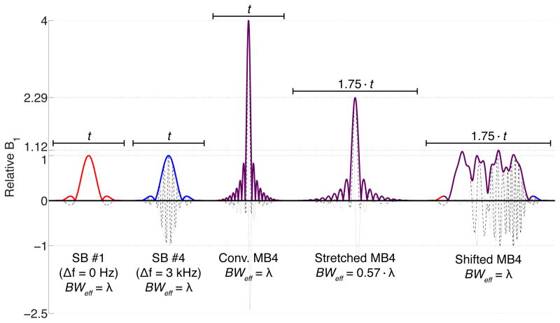
Purpose: To evaluate an alternative method for generating multibanded radiofrequency (RF) pulses for use in multiband slice-accelerated imaging with slice-GRAPPA unaliasing, substantially reducing the required peak power without bandwidth compromises. This allows much higher accelerations for spin-echo methods such as SE-fMRI and diffusion-weighted MRI where multibanded slice acceleration has been limited by available peak power.

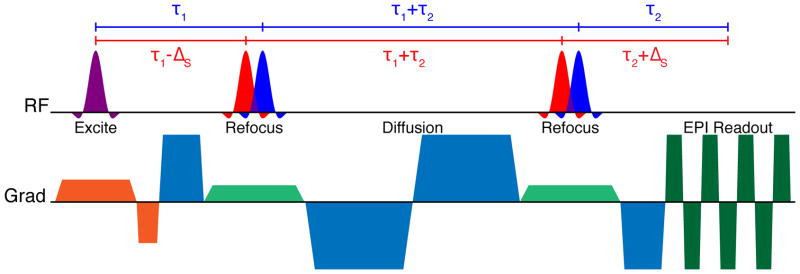
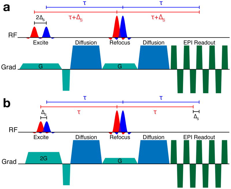
Theory and methods: Multibanded "time-shifted" RF pulses were generated by inserting temporal shifts between the applications of RF energy for individual bands, avoiding worst-case constructive interferences. Slice profiles and images in phantoms and human subjects were acquired at 3 T.

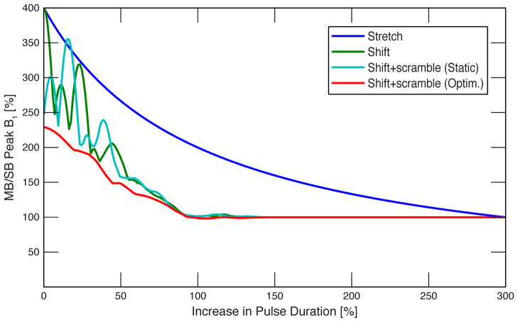
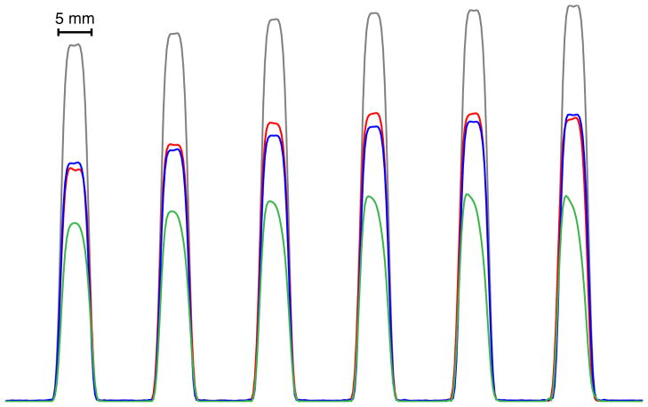
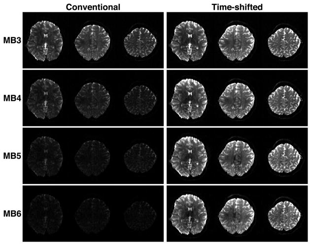
Results: For typical sinc pulses, time-shifted multibanded RF pulses were generated with little increase in required peak power compared to single-banded pulses. Slice profile quality was improved by allowing for higher pulse bandwidths, and image quality was improved by allowing for optimum flip angles to be achieved.

Conclusion: A simple approach has been demonstrated that significantly alleviates the restrictions imposed on achievable slice acceleration factors in multiband spin-echo imaging due to the power requirements of multibanded RF pulses. This solution will allow for increased accelerations in diffusion-weighted MRI applications where data acquisition times are normally very long and the ability to accelerate is extremely valuable.


Figures

Figure 1
Figure 1
From left to right: single-banded (SB) sinc RF pulse (R = 5.2) for band #1 (no frequency offset), SB pulse for band #4 (~3 kHz frequency offset), conventional four-banded (MB4) pulse with matched duration t, conventional MB4 pulse with stretched duration 1.75·t, time-shifted MB4 pulse generated with the proposed technique with duration 1.75·t (25% temporal shift between bands). For all plots, solid lines represent the magnitude, dashed lines the real component, and dotted lines the imaginary component.
Figure 2
Figure 2
Bipolar diffusion-weighted EPI sequence diagram using time-shifted multi-banded refocusing RF pulses. No time shift is used for the multi-banded excitation RF pulse.
Figure 3
Figure 3
Monopolar diffusion-weighted EPI sequences using time-shifted multi-banded refocusing and excitation RF pulses. The top diagram (a) shows the “aligned-echo” variant, where all spin echoes form at the same time, but the effective TE of each band differs by 2ΔS. The bottom diagram (b) shows the “aligned-TE” variant, where the TE of each band is the same, but the formation of the spin echoes for each band is separated in time by ΔS.
Figure 4
Figure 4
Plot of required peak B1 vs. total pulse duration for four-banded pulses with 1.25 kHz inter-band frequency offsets: stretched conventional pulse (dark blue), time-shifted pulse (green), time-shifted with static inter-band phase offsets (light blue), and time-shifted with optimized phase offsets for each shift (red). B1 and duration are shown relative to the base single-banded sinc pulse.
Figure 5
Figure 5
Slice profiles measured in a cylindrical oil phantom. For reference, slice profiles acquired separately with single-band pulses at the minimum TE of 28 ms are shown in gray. The six slices simultaneously acquired with a time-shifted six-banded RF pulse are shown in red (TE = 38 ms); this is compared to single-band acquisition at the same TE is shown in blue. The same six slices acquired with a stretched conventional six-banded RF pulse are shown in green (TE = 52 ms).
Figure 6
Figure 6
Comparison of human brain images acquired with multi-band slice accelerations of 3–6 (MB3-MB6) using conventional multi-banded RF pulses (left column) and time-shifted RF pulses (right column) with equivalent effective bandwidth. The display window and level are the same for all images.

References

    1. Feinberg DA, Moeller S, Smith SM, Auerbach EJ, Ramanna S, Glasser MF, Miller KL, Uğurbil K, Yacoub E. Multiplexed echo planar imaging for sub-second whole brain FMRI and fast diffusion imaging. PLoS ONE. 2010;5(12):e15710. - PMC - PubMed
    1. Koopmans PJ, Boyacioğlu R, Barth M, Norris DG. Whole brain, high resolution spin-echo resting state fMRI using PINS multiplexing at 7 T. Neuroimage. 2012;62(3):1939–1946. - PubMed
    1. Larkman DJ, Hajnal JV, Herlihy AH, Coutts GA, Young IR, Ehnholm G. Use of multicoil arrays for separation of signal from multiple slices simultaneously excited. J Magn Reson Imaging. 2001;13(2):313–317. - PubMed
    1. Moeller S, Auerbach E, van de Moortele P-F, Adriany G, Uğurbil K. fMRI with 16 fold reduction using multibanded multislice sampling. Proceedings of the 16th Annual Meeting of ISMRM; Toronto, Ontario, Canada. 2008. p. 2366.
    1. Moeller S, Yacoub E, Olman CA, Auerbach EJ, Strupp J, Harel N, Uğurbil K. Multiband multislice GE-EPI at 7 tesla, with 16-fold acceleration using partial parallel imaging with application to high spatial and temporal whole-brain fMRI. Magn Reson Med. 2010;63(5):1144–1153. - PMC - PubMed

Publication types

MeSH terms