. 2013 Mar 15;27(6):602-14.
doi: 10.1101/gad.213314.113. Epub 2013 Mar 6.

The BEN domain is a novel sequence-specific DNA-binding domain conserved in neural transcriptional repressors

Affiliations

The BEN domain is a novel sequence-specific DNA-binding domain conserved in neural transcriptional repressors

Qi Dai et al. Genes Dev. .

Abstract

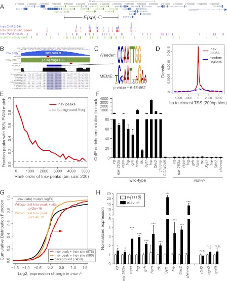
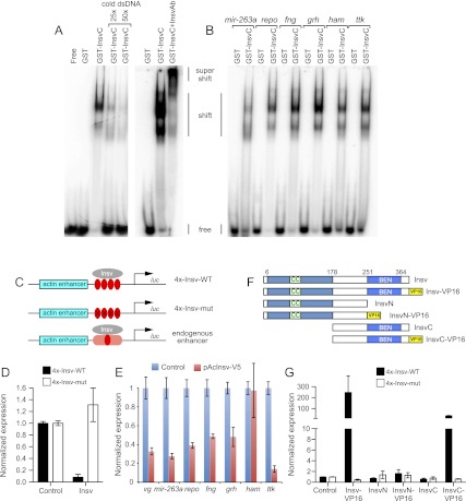
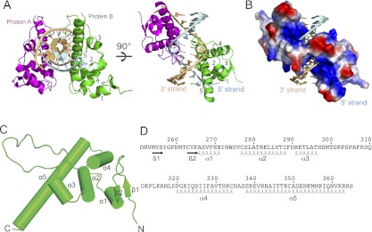
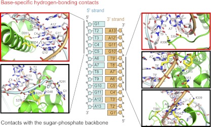
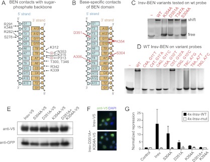
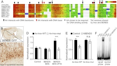
We recently reported that Drosophila Insensitive (Insv) promotes sensory organ development and has activity as a nuclear corepressor for the Notch transcription factor Suppressor of Hairless [Su(H)]. Insv lacks domains of known biochemical function but contains a single BEN domain (i.e., a "BEN-solo" protein). Our chromatin immunoprecipitation (ChIP) sequencing (ChIP-seq) analysis confirmed binding of Insensitive to Su(H) target genes in the Enhancer of split gene complex [E(spl)-C]; however, de novo motif analysis revealed a novel site strongly enriched in Insv peaks (TCYAATHRGAA). We validate binding of endogenous Insv to genomic regions bearing such sites, whose associated genes are enriched for neural functions and are functionally repressed by Insv. Unexpectedly, we found that the Insv BEN domain binds specifically to this sequence motif and that Insv directly regulates transcription via this motif. We determined the crystal structure of the BEN-DNA target complex, revealing homodimeric binding of the BEN domain and extensive nucleotide contacts via α helices and a C-terminal loop. Point mutations in key DNA-contacting residues severely impair DNA binding in vitro and capacity for transcriptional regulation in vivo. We further demonstrate DNA-binding and repression activities by the mammalian neural BEN-solo protein BEND5. Altogether, we define novel DNA-binding activity in a conserved family of transcriptional repressors, opening a molecular window on this extensive gene family.


Figures

Figure 1.
Figure 1.
ChIP-seq revealed a novel motif associated with transcriptional repression by Drosophila Insv. (A) Insv exhibits extensive binding to N/Su(H)-regulated enhancers throughout the E(spl)-C (green highlighted genes). (B) Insv binding to the fringe promoter. University of California at Santa Cruz (UCSC) Genome Browser view shows raw ChIP-seq signals from 2.5- to 6.5-h (“early,” blue track) and 6.5- to 12-h (“late,” green track) embryos; the processed peak locations are marked with red boxes. These peaks coincide with a well-conserved palindromic motif. (C) De novo motif analysis from both early and late data sets defines related motifs that are highly enriched among Insv-bound regions. (D) Insv peaks that bear this motif are strongly enriched near annotated transcription start sites (red line). The dashed blue line shows distances from 100,000 random genomic regions to the closest transcription starts. This analysis is from the early time point, but the same is true for the later time point (Supplemental Fig. 2) (χ2P-value < 2.2 × 10⁻¹⁶). (E) The Insv motif is preferentially localized in stronger Insv peaks in both early (here) and late embryo (Supplemental Fig. 1) time points. The dashed gray line shows the estimated background frequency of this site; the distributions are significantly different. (F) ChIP-qPCR validates binding of Insv to target genes bearing this motif; absence of signals in insv−/− embryos demonstrates specificity. (G) Cumulative distribution function of gene expression in 6- to 8-h wild-type and insv−/− embryos. Genes associated with Insv ChIP-seq peaks bearing the identified motif (red line) were generally up-regulated in insv mutants relative to background genes (black line), whereas genes bound by Insv but lacking the motif were not up-regulated (orange line); P-values from Wilcoxon rank sum test. (H) qPCR validation of the up-regulation of Insv target genes in insv mutant embryos. (*) P < 0.001; (***) P < 0.0001.
Figure 2.
Figure 2.
Insv is a novel DNA-binding protein that directly mediates transcriptional repression. (A) Gel shift analysis with radiolabeled probes demonstrates specific binding of the Insv BEN domain to a Insv site in the gene vg but not a mutated version. The shifted complexes can be competed away with cold probe and supershifted with Insv antibodies. (B) Insv binds to endogenous sites from a variety of neural target genes. (C) Schematics of actin-luciferase reporter constructs into which either wild-type or mutant Insv motifs or endogenous insv-bound genomic regions have been inserted. (D) Insv-binding sites directly recruit Insv for transcriptional repression. Actin-luciferase reporters containing multimers of Insv-binding sites are repressed upon cotransfection of Insv plasmid relative to empty plasmid; no effects were observed with a similar reporter bearing point mutations in the Insv site. (E) Endogenous Insv-bound sites from a variety of neural target genes were similarly repressed. (F) Insv variant proteins used for structure–function tests. (G) Fusion of the VP16 activation domain to Insv or InsvC converts them into transcriptional activators; the latter demonstrates that DNA-binding activity localizes to the BEN domain.
Figure 3.
Figure 3.
Overall structure of the Insv-BEN domain in the DNA-bound state. (A) Two views of the structure of the complex containing two BEN domains (in ribbon representation) bound to a self-complementary 13-mer DNA duplex (strands colored in light blue and gold). The symmetry-related Ben domains are colored in green and magenta, with α helices numbered from 1 to 5. (B) The same view of the complex as in the right panel of A, with protein in an electrostatic surface representation. (C) Helical topology of the Insv Ben domain in complex with its DNA target site. Ribbon representation of the Insv BEN domain reveals a predominantly α-helical structure. (D) Positioning of α helices and β strands within the Insv BEN domain.
Figure 4.
Figure 4.
Amino acid contacts of the BEN domain with the DNA backbone and specific nucleotide side chains. Shown are details of intermolecular hydrogen-bonding contacts in the BEN–DNA complex. The sequence and numbering scheme are shown in the middle panel. Intermolecular hydrogen-bonding contacts involving the G1-T2-T3-C4-C5 segment on the 5′ strand are shown in the panels on the left, while those involving contacts with the C5′-A6′-A7′-T8′-T9′ of the 3′ strand are shown on the right. Base-specific hydrogen-bonding contacts are shown in red boxes, while those involving contacts with the sugar-phosphate backbone are shown in black boxes.
Figure 5.
Figure 5.
Functional assays confirm the structure of the BEN:DNA complex. (A) Summary of intermolecular hydrogen-bonding contacts to the sugar-phosphate backbone. (B) Summary of base-specific intermolecular hydrogen-bonding contacts. (C) Mutation of several BEN domain residues involved in base-specific recognition abrogates their functionality in gel shift assays visualized with ethidium bromide. (D) Mutations of DNA base pairs involved in base-specific recognition by the BEN domain abrogate association with Insv-BEN in gel shift assays. (E) A panel of V5-tagged Insv proteins bearing alanine mutations of side chain-contacting residues in the BEN domain were transfected into S2 cells. Western analysis showed normal accumulation of all of the Insv variants. (F) Immunostaining of Insv constructs in S2 cells showed that both wild type and the D351A+K354A double mutant exhibited nuclear localization (as did other mutant Insv proteins) (see Supplemental Fig. 5). (G) BEN domain mutants of Insv were cotransfected with wild-type and mutant insv transcriptional reporters. S304A had only mild effects, but D351A and K354A were strongly compromised for transcriptional repression. The D351A+K354A double mutant was completely inactive.
Figure 6.
Figure 6.
Conservation of DNA-binding capacity in a mammalian neural BEN-solo protein. (A) Alignment of the Insv BEN domain (starting with the second α helix) (see Fig. 3) with BEN domains from other proteins reveals conservation of critical DNA-contacting residues in some Drosophila and mammalian proteins. (B) A BAC transgenic mouse bearing a knock-in of GFP into BEND5 exhibits specific expression in the cortex (data from the GENSAT project, http://www.gensat.org). The boxed region is enlarged in C and highlights expression of BEND5 in layer V pyramidal neurons. (D) A VP16 fusion to the BEN domain of human BEND5 can activate the Insv reporter but does not affect the mutant reporter. (E) Full-length human BEND5 represses a wild-type but not mutant Insv reporter in HEK293T cells. (F) Gel shift analysis using radiolabeled probes confirms specific binding of hBEND5 to a probe from the Drosophila vg gene bearing an Insv site but not a vg probe mutated at the Insv-binding site.

References

    1. Abhiman S, Iyer LM, Aravind L 2008. BEN: A novel domain in chromatin factors and DNA viral proteins. Bioinformatics 24: 458–461 - PMC - PubMed
    1. Adams PD, Grosse-Kunstleve RW, Hung LW, Ioerger TR, McCoy AJ, Moriarty NW, Read RJ, Sacchettini JC, Sauter NK, Terwilliger TC 2002. PHENIX: Building new software for automated crystallographic structure determination. Acta Crystallogr D Biol Crystallogr 58: 1948–1954 - PubMed
    1. Aoki T, Sarkeshik A, Yates J, Schedl P 2012. Elba, a novel developmentally regulated chromatin boundary factor is a hetero-tripartite DNA binding complex. eLife 1: e00171. - PMC - PubMed
    1. Ashburner M, Ball CA, Blake JA, Botstein D, Butler H, Cherry JM, Davis AP, Dolinski K, Dwight SS, Eppig JT, et al. 2000. Gene ontology: Tool for the unification of biology. The Gene Ontology Consortium. Nat Genet 25: 25–29 - PMC - PubMed
    1. Bailey TL, Elkan C 1994. Fitting a mixture model by expectation maximization to discover motifs in biopolymers. Proc Int Conf Intell Syst Mol Biol 2: 28–36 - PubMed

Publication types

MeSH terms