. 2013 May;87(10):5564-76.
doi: 10.1128/JVI.03426-12. Epub 2013 Mar 6.

Effects of postchallenge administration of ST-246 on dissemination of IHD-J-Luc vaccinia virus in normal mice and in immune-deficient mice reconstituted with T cells

Affiliations

Effects of postchallenge administration of ST-246 on dissemination of IHD-J-Luc vaccinia virus in normal mice and in immune-deficient mice reconstituted with T cells

Marina Zaitseva et al. J Virol. 2013 May.

Abstract

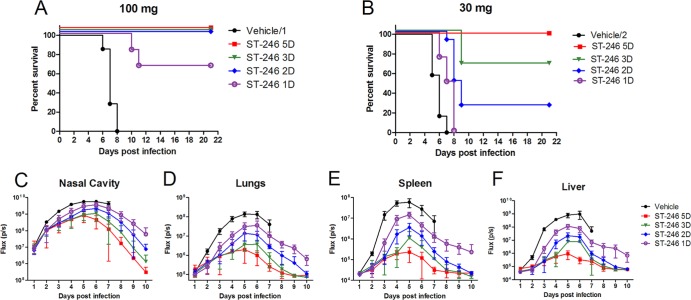
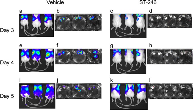
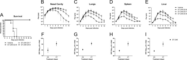
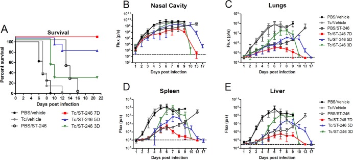
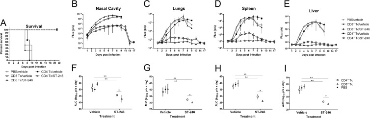
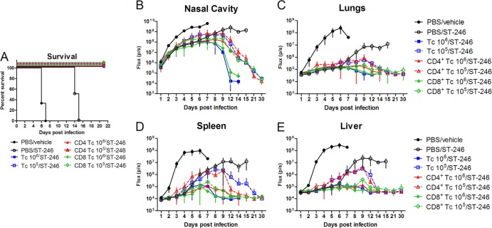
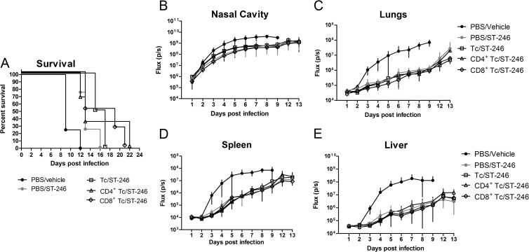
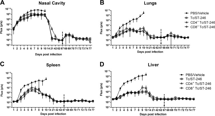
Whole-body bioimaging was used to study dissemination of vaccinia virus (VACV) in normal and in immune deficient (nu(-)/nu(-)) mice protected from lethality by postchallenge administration of ST-246. Total fluxes were recorded in the liver, spleen, lungs, and nasal cavities of live mice after intranasal infection with a recombinant IHD-J-Luc VACV expressing luciferase. Areas under the flux curve were calculated for individual mice to assess viral loads. Treatment for 2 to 5 days of normal BALB/c mice with ST-246 at 100 mg/kg starting 24 h postchallenge conferred 100% protection and reduced viral loads in four organs compared to control mice. Mice also survived after 5 days of treatment with ST-246 at 30 mg/kg, and yet the viral loads and poxes were higher in these mice compared to 100-mg/kg treatment group. Nude mice were not protected by ST-246 alone or by 10 million adoptively transferred T cells. In contrast, nude mice that received T cells and 7-day treatment with ST-246 survived infection and exhibited reduced viral loads compared to nonreconstituted and ST-246-treated mice after ST-246 was stopped. Similar protection of nude mice was achieved using adoptively transferred 1.0 and 0.1 million, but not 0.01 million, purified T cells or CD4(+) or CD8(+) T cells in conjunction with ST-246 treatment. These data suggest that ST-246 protects immunocompetent mice from lethality and reduces viral dissemination in internal organs and poxvirus lesions. Furthermore, immune-deficient animals with partial T cell reconstitution can control virus replication after a course of ST-246 and survive lethal vaccinia virus challenge.


Figures

Fig 1
Fig 1
Two days of treatment with ST-246 at 100 mg/kg dose starting 24 h postchallenge with IHD-J-Luc VACV was required to protect 100% of BALB/c mice from lethality. (A to F) Lethality outcome and bioluminescence measurements in the internal organs of BALB/c mice infected with recombinant IHD-J-Luc VACV. BALB/c mice were infected intranasally with IHD-J-Luc VACV at 105 PFU. Starting 24 h postchallenge, mice received daily treatments with vehicle alone for 5 days (black circles) or with ST-246 for 1 day (purple open circle), 2 days (blue diamond), 3 days (green triangle), or for 5 days (red square) (A to F) at 100 mg/kg (A, C to F) or at 30 mg/kg (B). Mice were observed for survival (A and B) and were subjected to whole-body bioimaging (C to F). Total fluxes in the nasal cavity (B), lungs (D), liver (E), and spleen (F) were determined and used to calculate the mean total flux ± the standard deviation (SD) using a Student t test. Vehicle/1, 28 animals per group; vehicle/2, 18 animals per group; all ST-246-treated mice, 6 mice per group.
Fig 2
Fig 2
ST-246 treatment prevented dissemination of IHD-J-Luc VACV into the ovaries of normal BALB/c mice. BALB/c mice (9 mice per group) were infected with IHD-J-Luc and were treated daily for 3 days with vehicle (a, b, e, f, i, and j) or with a full dose of ST-246 (c, d, g, h, k, and l). On days 3 (a to d), 4 (e to h), and 5 (i to l), 3 mice per group were imaged and, immediately after imaging, mice were sacrificed, and the ovaries were imaged ex vivo in the wells of a 24-well plate. Dorsal images of live mice and of extracted ovaries (two ovaries from one mouse in a single well) are shown in panels a, c, e, g, i, and k and in panels b, d, f, h, j, and l, respectively.
Fig 3
Fig 3
ST-246 started 2 or 3 days, but not 4 days, postinfection protected mice from lethal challenge with IHD-J-Luc VACV. BALB/c mice were infected with IHD-J-Luc as described in Fig. 1 and were treated for a total of 5 days with vehicle on days 2 to 6 (●) or with ST-246 at 100 mg/kg on days 2 to 6 (♢), days 3 to 7 (△), or days 4 to 8 (□) (A to E). Mice were observed for lethality (A) and were subjected to bioimaging daily for 10 days (B to E). Total fluxes in the nasal cavity (B), lungs (C), spleen (D), and liver (E) were determined and used to calculate the mean total flux ± the SD using a Student t test. (F to I) AUCs were calculated for fluxes from individual mice for days 1 to 10 in the nasal cavities (F), lungs (G), spleens (H), and livers (I) of mice that received ST-246 on day 2 to 6 and days 3 to 7 only. The data are shown as mean AUCs ± 2×SE for groups of 6 mice per group. The experiment was performed twice with similar results.
Fig 4
Fig 4
Nude mice reconstituted with 10 million T cells were protected from lethality when they received ST-246 for no less than 1 week. Nude mice received an adoptive transfer of 10 million purified T cells, were infected with IHD-J-Luc VACV at 104 PFU, and were treated daily with vehicle alone for 7 days (gray circles), or with ST-246 at 100 mg/kg for 7 days (red square), 5 days (blue triangle), or 3 days (inverted green triangle). In control, nude mice received transfer of PBS (sham adoptive transfer), were infected, and were treated daily for 7 days with vehicle alone (black circles) or with ST-246 (black open circles). Mice were observed for lethality (A) and were subjected to bioimaging daily to calculate mean fluxes ± the SD in the nasal cavity (B), lungs (C), spleen (D), and liver (E). PBS/vehicle, Tc/vehicle, Tc/ST-246 5D, and Tc/ST-246 3D, 8 mice per group; PBS/ST-246 and Tc/ST-246 7D, 4 mice per group. Mean background levels of fluxes (p/s, photons per second) ± the SD were recorded in nude mice prior to infection in the nasal cavity (23.3 × 10³ ± 4.5 × 10³), lungs (36.3 × 10³ ± 11.6 × 10³), spleen (10.9 × 10³ ± 2.5 × 10³), and liver (30.2 × 10³ ± 2.1 × 10³) and are shown as broken lines (means only) in panels B, C, D, and E, respectively. The experiment was performed twice with similar results.
Fig 5
Fig 5
Adoptively transferred CD4+ and CD8+ T cells conferred protection from lethality and reduced viral loads in the recipient nude mice after ST-246 treatment. Nude mice received an adoptive transfer with 10 million of CD8+ T cells (closed and open triangles) or CD4+ T cells (closed and open squares) or were sham transferred (gray circles) (A to E). All mice were infected with 104 PFU of IHD-J-Luc and were treated daily with ST-246 at 100 mg/kg (closed triangles and closed squares) or with equal volume of vehicle (gray circles, open triangles, and open squares) for 7 days starting 24 h postchallenge (A to E). Mice were observed for lethality (A) and were subjected to bioimaging (B to E). Total fluxes in the nasal cavity (B), lungs (C), spleen (D), and liver (E) were determined and used to calculate mean total flux ± the SD using a Student t test. (F to I) AUCs were calculated for fluxes from individual mice for first 7 days in the nasal cavities (F), lungs (G), spleens (G), and livers (H) and used to calculate mean AUCs ± 2×SE for mice that received sham transfer with PBS (black circles) or were reconstituted with CD4+ (open squares) or CD8+ T cells (open triangles). Significant differences are shown between CD4+ or CD8+ T cell-reconstituted and ST-246-treated mice and sham-reconstituted vehicle-treated mice and for ST-246-treated nude mice reconstituted with CD4+ versus CD8+ T cells. *, P ≤ 0.05; **, P ≤ 0.001. PBS/vehicle, CD8+ Tc/ST-246, 4 mice per group; CD4+ Tc/vehicle and CD8+ Tc/vehicle, 3 mice per group; CD4+ Tc/ST-246, 5 mice per group. The experiment was performed twice with similar results.
Fig 6
Fig 6
One million and 0.1 million of T cells transferred to nude mice were sufficient to protect from lethality in conjunction with 7-day treatment with ST-246. One day prior to infection with 104 PFU of IHD-J-Luc, nude mice received an adoptive transfer with 1.0 or 0.1 million of T cells (blue closed and open squares) or of CD4+ (red closed and open triangles) or CD8+ T cells (green closed and open diamonds) from normal BALB/c mice and starting 1 day postinfection, were all treated with ST-246 for 7 days. Control mice were sham transferred with PBS 1 day before infection and after infection received vehicle or ST-246 (black closed and open circles) for 7 days. Mice were observed for lethality (A) and were subjected to bioimaging until day 14 or 15 for all surviving mice and additionally on days 21 and 30 for the group of mice that were reconstituted with 0.1 million of T cells or T cell subsets to confirm complete clearance of IHD-J-Luc from all organs. Recorded fluxes were used to calculate mean fluxes ± the SD in the nasal cavity (B), lungs (C), spleen (D), and liver (E). PBS/vehicle, 3 mice per group; PBS/ST-246, Tc 106/ST-246, and Tc 105/ST-246, 4 mice per group. The experiment was performed twice with similar results.
Fig 7
Fig 7
Ten thousand T cells or CD4+ or CD8+ T cells transferred into nude mice did not protect from lethality and from viral dissemination. Nude mice received a sham transfer with PBS (black and gray circles) or with 104 T cells (open squares), CD4+ T cells (open triangles), or CD8+ T cells (open diamonds). All mice were infected with 104 PFU of IHD-J-Luc 1 day after reconstitution and treated with vehicle (black circles) or with ST-246 (gray circles and open symbols) daily for 7 days starting 24 h postinfection. Mice were observed for lethality (A) and were subjected to bioimaging daily to calculate mean fluxes ± the SD in the nasal cavity (B), lungs (C), spleen (D), and liver (E). Four mice per group were used in the experiment.
Fig 8
Fig 8
Nude mice reconstituted with 0.1 million of T cells or of CD4+ or CD8+ T cells were protected from rechallenge with IHD-J-Luc without the need for additional ST-246 treatment. Nude mice were sham transferred with PBS (●) or received adoptive transfer with 0.1 million of T cells (□) or of CD4+ (△) or CD8+ T cells (♢) and were infected with 104 PFU IHD-J-Luc 1 day after reconstitution. Sham-transferred mice received vehicle (PBS/vehicle) and T cell reconstituted mice received ST-246 for 7 days (Tc/ST-246, CD4+ Tc/ST-246, and CD8+ Tc/ST-246). On day 67 postinfection, all survived mice were rechallenged with 104 PFU of IHD-J-Luc. Mice were imaged after primary infection up to day 62 at time points indicated on the x axis and then daily for 10 days after rechallenge. Recorded fluxes were used to calculate mean fluxes ± the SD in the nasal cavity (A), lungs (B), spleen (C), and liver (D). Vertical arrow indicates time of rechallenge on day 67. Four mice per group were used in all groups. The experiment was performed twice with similar results.

References

    1. Henderson DA, Inglesby TV, Bartlett JG, Ascher MS, Eitzen E, Jahrling PB, Hauer J, Layton M, McDade J, Osterholm MT, O'Toole T, Parker G, Perl T, Russell PK, Tonat K. 1999. Smallpox as a biological weapon: medical and public health management. JAMA 281:2127–2137 - PubMed
    1. Rimoin AW, Mulembakani PM, Johnston SC, Lloyd Smith JO, Kisalu NK, Kinkela TL, Blumberg S, Thomassen HA, Pike BL, Fair JN, Wolfe ND, Shongo RL, Graham BS, Formenty P, Okitolonda E, Hensley LE, Meyer H, Wright LL, Muyembe JJ. 2010. Major increase in human monkeypox incidence 30 years after smallpox vaccination campaigns cease in the Democratic Republic of Congo. Proc. Natl. Acad. Sci. U. S. A. 107:16262–16267 - PMC - PubMed
    1. Whitley RJ. 2003. Smallpox: a potential agent of bioterrorism. Antivir. Res. 57:7–12 - PubMed
    1. Amanna IJ, Slifka MK, Crotty S. 2006. Immunity and immunological memory following smallpox vaccination. Immunol. Rev. 211:320–337 - PubMed
    1. Hammarlund E, Lewis MW, Hansen SG, Strelow LI, Nelson JA, Sexton GJ, Hanifin JM, Slifka MK. 2003. Duration of antiviral immunity after smallpox vaccination. Nat. Med. 9:1131–1137 - PubMed

Publication types

MeSH terms

LinkOut - more resources