. 2013;9(2):e1003334.
doi: 10.1371/journal.pgen.1003334. Epub 2013 Feb 28.

The amidation step of diphthamide biosynthesis in yeast requires DPH6, a gene identified through mining the DPH1-DPH5 interaction network

Affiliations

The amidation step of diphthamide biosynthesis in yeast requires DPH6, a gene identified through mining the DPH1-DPH5 interaction network

Shanow Uthman et al. PLoS Genet. 2013.

Abstract

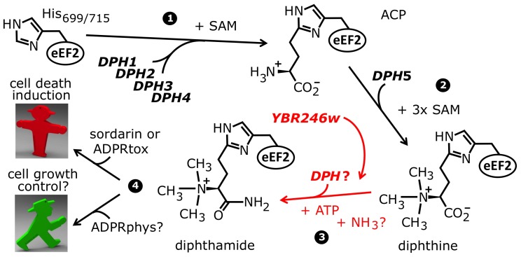
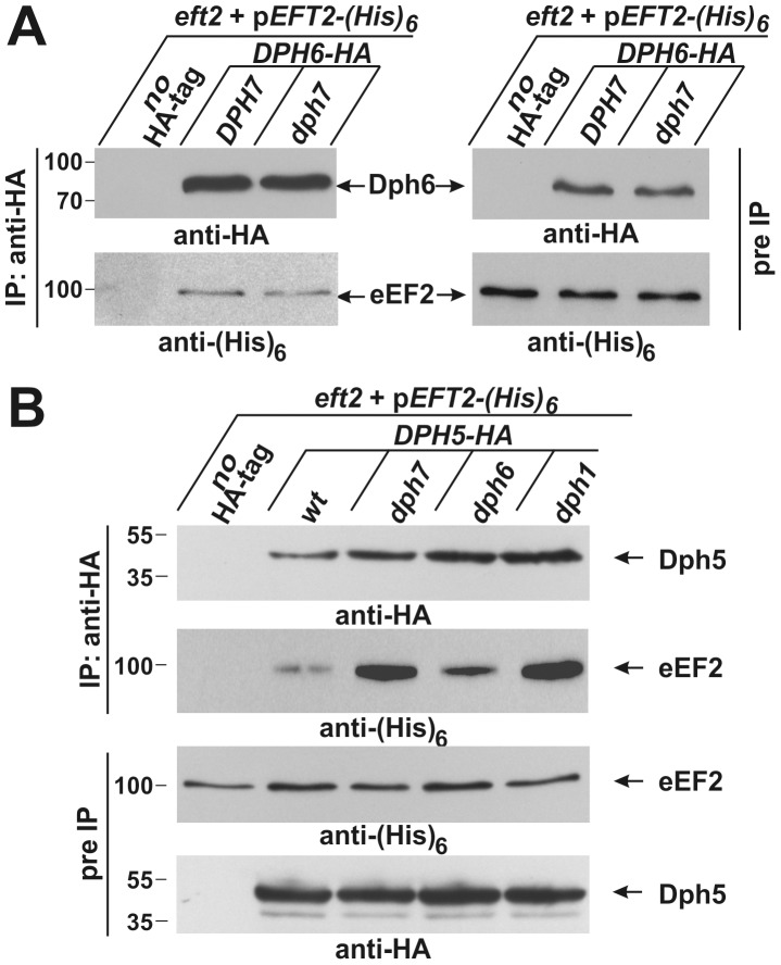
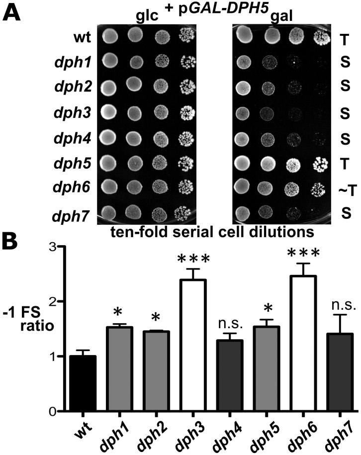
Diphthamide is a highly modified histidine residue in eukaryal translation elongation factor 2 (eEF2) that is the target for irreversible ADP ribosylation by diphtheria toxin (DT). In Saccharomyces cerevisiae, the initial steps of diphthamide biosynthesis are well characterized and require the DPH1-DPH5 genes. However, the last pathway step-amidation of the intermediate diphthine to diphthamide-is ill-defined. Here we mine the genetic interaction landscapes of DPH1-DPH5 to identify a candidate gene for the elusive amidase (YLR143w/DPH6) and confirm involvement of a second gene (YBR246w/DPH7) in the amidation step. Like dph1-dph5, dph6 and dph7 mutants maintain eEF2 forms that evade inhibition by DT and sordarin, a diphthamide-dependent antifungal. Moreover, mass spectrometry shows that dph6 and dph7 mutants specifically accumulate diphthine-modified eEF2, demonstrating failure to complete the final amidation step. Consistent with an expected requirement for ATP in diphthine amidation, Dph6 contains an essential adenine nucleotide hydrolase domain and binds to eEF2. Dph6 is therefore a candidate for the elusive amidase, while Dph7 apparently couples diphthine synthase (Dph5) to diphthine amidation. The latter conclusion is based on our observation that dph7 mutants show drastically upregulated interaction between Dph5 and eEF2, indicating that their association is kept in check by Dph7. Physiologically, completion of diphthamide synthesis is required for optimal translational accuracy and cell growth, as indicated by shared traits among the dph mutants including increased ribosomal -1 frameshifting and altered responses to translation inhibitors. Through identification of Dph6 and Dph7 as components required for the amidation step of the diphthamide pathway, our work paves the way for a detailed mechanistic understanding of diphthamide formation.


Conflict of interest statement

The authors have declared that no competing interests exist.

Figures

Figure 1
Figure 1. The biosynthetic pathway for modification of eEF2 by diphthamide.
For roles played by the bona fide diphthamide genes DPH1–DPH5 in steps 1 and 2 of the pathway, see main text. The ill-defined step 3, conversion of diphthine to diphthamide by amidation, is highlighted (red label). It likely involves ATP and ammonium cofactors in a reaction catalyzed by unidentified DPH gene product(s). Step 4 indicates diphthamide can be hijacked for eEF2 inactivation and cell death induction by antifungals, i.e. sordarin and bacterial ADP ribosylase toxins (ADPRtox); alternatively, it has been reported to undergo cell growth related physiological ADP ribosylation (ADPRphys?) by elusive cellular modifier(s). ACP, 2-[3-amino-carboxyl-propyl]-histidine; SAM: S-adenosylmethionine.
Figure 2
Figure 2. Genome-wide gene interaction databases identify additional diphthamide related candidate genes: YLR143w/DPH6 and YBR246w/DPH7.
(A) SGA database (DRYGIN). Genetic interaction profiles among DPH1, DPH2, DPH4, DPH5, YBR246w and YLR143w query gene deletion strains and 3885 or 4457 array ORF mutants were extracted from data for a total of ∼1700 query strains deposited at DRYGIN (for full details, see excel spread sheet in Table S1). Ranking of top interactors for each query ORF was according to PCC (Pearson correlation coefficient) determination. For simplicity, array ORFs DPH1, DPH2, DPH4, DPH5, EFT1, EFT2 (shown in bold) as well as potentially diphthamide related candidate loci YLR143w and YBR246w (red circles) are listed that score repeatedly as significantly high interactors of the query ORFs. (B) Yeast Fitness database (FitDB). Genes whose deletions phenocluster with the six query ORFs above were extracted from FitDB, which is based on genome-scale co-fitness defect analysis of homozygous yeast deletion mutants in response to greater than 1144 different conditions. For simplicity, the top ten interactors for three of the six query genes (DPH5, YLR143w and YBR246w: pale blue central nodes) above are depicted. (C) Representation of the tightly clustered and expanded DPH1-DPH7 gene network where nodes (pale blue) correspond to individual DPH gene family members and edges connect gene pairs by PCC>0.14. Enhanced gene interaction strength is proportional to PCC stringency. Enriched GO process likelihoods in the diphthamide modification pathway are listed as P-values for the identified candidates DPH6/YLR143w and DPH7/YBR246w.
Figure 3
Figure 3. DPH6 and DPH7 deletion strains copy traits typically related to the bona fide diphthamide mutants dph1-dph5.
(A) Sordarin resistance. Ten-fold serial cell dilutions of the indicated yeast strains, BY4741 wild-type (wt) background and its dph1-dph7 gene deletion derivatives (upper panels) as well an MKK-derived eft1 eft2 double deletion background maintaining plasmid pEFT2 wild-type or H699 substitution (H699 N and H699I) alleles of EFT2 (lower panels), were grown on YPD plates in the absence (control) or presence (+sor) of 10 µg ml−1 sordarin. Growth was assayed for 3 d at 30°C. Sordarin resistant (R) and sensitive (S) responses are indicated. (B) Lack of in vitro ADP ribose acceptor activity of eEF2. Cell extracts obtained from dph1, dph5, dph6 and dph7 mutant and wild-type (wt) strains were incubated with (+DT) or without (−DT) 20 nM diphtheria toxin in the presence of biotin-NAD (10 µM) at 37°C for 1 hour. The transfer of biotin-ADP-ribose to eEF2 was detected by Western blotting using a streptavidin-conjugate. Two unknown non-specific bands (indicated by *) served as internal controls for even sample loading. (C) DT phenotype. As indicated, yeast dph mutants and wild-type control (wt) were tested for sensitivity to intracellular expression of DTA, the cytotoxic ADP ribosylase fragment of DT. This in vivo assay involved galactose-inducible expression from vector pSU8 (see Materials and Methods). Serial cell dilutions were replica spotted onto raffinose (2% raf) and galactose-inducing conditions using concentrations (2, 0.2 and 0.1% gal) suited to achieve gradual down-regulation of DTA toxicity. Growth was for 3 days at 30°C. DTA sensitive (S) resistant (R), partially resistant (PR) and reduced sensitive (RS) phenotypes are indicated.
Figure 4
Figure 4. MS/MS spectra of diphthamide-, ACP-, and diphthine-modified EF2 peptide 686-VNILDVTLHADAIHR-700 from wild-type and mutant yeast strains.
Spectra are shown for (A) diphthamide-modified peptide from the wild-type yeast strain; (B) ACP-modified peptide from the dph5Δ mutant; (C) diphthine-modified peptide in the dph7Δ strain; (D) diphthine-modified peptide in the dph6Δ strain; (E) diphthine-modified peptide in the dph6Δ strain with loss of the trimethylamino group before analysis in the mass spectrometer indicated by the parent ion m/z. In each case the parent ion m/z and charge state is indicated. In (A), (C) and (D), * indicates neutral loss of trimethylamino during MS/MS. The inset in (C) shows greater detail for the more crowded part of the MS/MS spectrum. Figure S2A indicates how the B and Y ions are derived from the peptide sequence.
Figure 5
Figure 5. Co-immune precipitations reveal eEF2 interactions with Dph6 and Dph5.
(A) eEF2 interacts with Dph6 in a fashion that is independent of Dph7. (B) eEF2 interaction with Dph5 is dramatically enhanced by elimination of Dph7 or Dph1. Yeast strains co-expressing (His)6-tagged eEF2 with Dph6-HA (A) or Dph5-HA (B) in the background of wild-type (A: DPH7 and B: wt) and dph mutant strains (A: dph7; B: dph1, dph6 and dph7) were subjected to immune precipitations (IP) using the anti-HA antibody. Strains expressing (His)6-tagged eEF2 on their own served as IP controls (A and B: no HA-tag). Subsequently, the precipitates were probed with anti-HA (A: top left panel; B: first panel) and anti-(His)6 antibodies (A: bottom left panel) to check for the content of Dph6-HA (A) and Dph5-HA (B), respectively (all indicated by arrows). The content of HA-tagged Dph6 (A) and Dph5 (B) as well as (His)6-marked eEF2 (A and B) in the protein extracts prior to IP (pre-IP) was examined on individual Western blots using anti-HA (A: top right panel; B: fourth panel) and anti-(His)6 antibodies (A: bottom right panel; B: third panel), respectively. While absence of Dph7 hardly affected the Dph6•eEF2 interaction (A), Dph5•eEF2 interaction was strongly enhanced by inactivating DPH7 or DPH1 (B).
Figure 6
Figure 6. dph mutants show sensitivity to elevated diphthine synthase levels and confer reduced translational accuracy.
(A) DPH5 overexpression in dph1-dph4 and dph7 mutants causes cytotoxicity and a severe cell growth defect. Cells of yeast strains with the indicated genetic backgrounds and maintaining plasmid pGAL-DPH5 for galactose inducible overexpression of diphthine synthase Dph5 were serially diluted and replica spotted onto glucose (2% glc) and galactose (2% gal) media to assay their response to DPH5 overexpression. Growth was for 3 days at 30°C. Unaltered (T), slightly weakened tolerance (∼T) and sensitive (S) responses are indicated. Note that dph1-dph4 and dph7 mutants are extremely sensitive to DPH5 overexpression. (B) Ribosomal frameshift analysis reveals erroneous translation in dph1-dph7 mutants. Strains with the indicated genetic backgrounds were transformed with control (pJD240.0) or lacZ −1 frameshift (pJD240.−1) plasmids to monitor lacZ expression through β-galactosidase (β-Gal) production using O-nitrophenol-D- galactopyranoside assays and to score translation efficiency (pJD240.0) and fidelity (pJD240.−1). Ribosomal −1 frameshifts are expressed relative to the level of overall translation efficiency with statistical significance determined by one-way ANOVA followed by Dunnett's multiple comparison. With the exception of dph4 and dph7, post-hoc comparison found that all other mutant backgrounds showed a significant increase in ribosomal −1 frameshifting relative to wild-type (wt) yeast cells (* = P<0.05; *** = P<0.001; ns. = not significant).
Figure 7
Figure 7. Both the Alpha_ANH_like_IV and YjgF-YER057c-UK114 domains in Dph6 are essential for its functionality.
(A) Diagram showing the DPH6 wild-type and mutant constructs tested in (B), indicating the Alpha_ANH_like_IV (ANH) and YjgF-YER057c-UK114 (UK114) domains and the position of point mutations, an in-frame deletion (- - - - -) and triple myc epitope tag (myc 3) as appropriate. (B) Ten-fold serial cell dilutions of a dph6 deletion strain carrying the constructs shown in (A) or the corresponding empty vector (top panel, pSU6 [wt DPH6]; lower panel, pSU7 [wt DPH6]: Table S3) were spotted onto SCD-Leu plates with or without 10 µg/ml sordarin and grown at 30°C for 3 days.
Figure 8
Figure 8. Model for the diphthamide pathway incorporating the proposed novel roles of Dph5, Dph6, and Dph7.
(A) Diphthamide pathway showing interaction of Dph5 with unmodified eEF2 and the proposed role of Dph7 in displacement of Dph5 prior to diphthine amidation. (B) Elimination of the trimethylamino group in the absence of the proposed amidase Dph6 suggesting lability of diphthine in its absence.

Comment in

References

    1. Ahrne E, Muller M, Lisacek F (2010) Unrestricted identification of modified proteins using MS/MS. Proteomics 10: 671–686. - PubMed
    1. Seet BT, Dikic I, Zhou MM, Pawson T (2006) Reading protein modifications with interaction domains. Nat Rev Mol Cell Biol 7: 473–483. - PubMed
    1. Walsh CT, Garneau-Tsodikova S, Gatto GJJ (2005) Protein posttranslational modifications: the chemistry of proteome diversifications. Angew Chem Int Ed Engl 44: 7342–7372. - PubMed
    1. Greganova E, Altmann M, Butikofer P (2011) Unique modifications of translation elongation factors. FEBS J 278: 2613–2624. - PubMed
    1. Van Ness BG, Howard JB, Bodley JW (1980) ADP-ribosylation of elongation factor 2 by diphtheria toxin. NMR spectra and proposed structures of ribosyl-diphthamide and its hydrolysis products. J Biol Chem 255: 10710–10716. - PubMed

Publication types

MeSH terms