. 2013;8(2):e56028.
doi: 10.1371/journal.pone.0056028. Epub 2013 Feb 26.

Interleukin-19 mediates tissue damage in murine ischemic acute kidney injury

Affiliations

Interleukin-19 mediates tissue damage in murine ischemic acute kidney injury

Yu-Hsiang Hsu et al. PLoS One. 2013.

Abstract

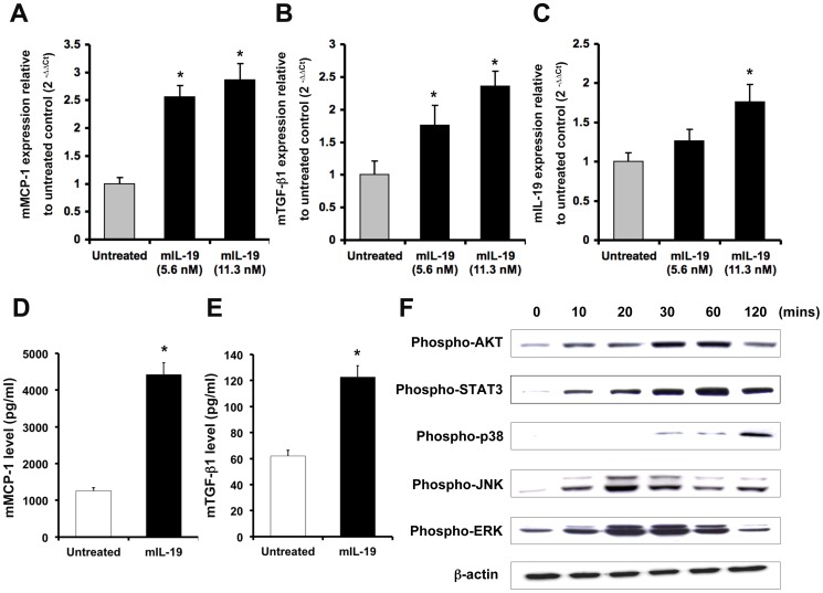
Inflammation and renal tubular injury are major features of acute kidney injury (AKI). Many cytokines and chemokines are released from injured tubular cells and acts as proinflammatory mediators. However, the role of IL-19 in the pathogenesis of AKI is not defined yet. In bilateral renal ischemia/reperfusion injury (IRI)-induced and HgCl2-induced AKI animal models, real-time quantitative (RTQ)-PCR showed that the kidneys, livers, and lungs of AKI mice expressed significantly higher IL-19 and its receptors than did sham control mice. Immunohistochemical staining showed that IL-19 and its receptors were strongly stained in the kidney, liver, and lung tissue of AKI mice. In vitro, IL-19 upregulated MCP-1, TGF-β1, and IL-19, and induced mitochondria-dependent apoptosis in murine renal tubular epithelial M-1 cells. IL-19 upregulated TNF-α and IL-10 in cultured HepG2 cells, and it increased IL-1β and TNF-α expression in cultured A549 cells. In vivo, after renal IRI or a nephrotoxic dose of HgCl2 treatment, IL-20R1-deficient mice (the deficiency blocks IL-19 signaling) showed lower levels of blood urea nitrogen (BUN) in serum and less tubular damage than did wild-type mice. Therefore, we conclude that IL-19 mediates kidney, liver, and lung tissue damage in murine AKI and that blocking IL-19 signaling may provide a potent therapeutic strategy for treating AKI.


Conflict of interest statement

Competing Interests: The authors have declared that no competing interests exist.

Figures

Figure 1
Figure 1. Higher expression of IL-19 and its receptors in ischemic AKI mice.
(A) Serum BUN levels and (B) Renal tubular damage were analyzed at the indicated times for monitor renal function in mice with IRI-induced AKI. *P<0.05 compared with the corresponding sham-operated mice. (C–E) Transcript levels of IL-19 and its receptors in the kidney tissue of ischemic AKI mice. Experimental mice were killed 2 days after renal IRI. RTQ-PCR was done with primers specific for IL-19, IL-20R1, and IL-20R2. The relative quantification of PCR products was expressed as 2−ΔΔCt, corrected using GAPDH expression, and relative to levels of untreated cells. Data are the means ± SD of three experiments. *P<0.05 compared with sham-operated mice.
Figure 2
Figure 2. Expression of IL-19 and its receptors in three vital organs of ischemic AKI mice.
(A) AKI mice (n = 5 in each group) were killed 48 h after renal IRI. Paraffined sections of kidney, liver, and lung tissue were stained using anti-IL-19 mAb, anti-IL-20R1 mAb, and anti-IL-20R2 polyclonal Ab. Anti-mIgG1 was a negative control. The reaction was detected using AEC chromogen stain (red), and the nuclei were counterstained with hematoxylin (blue). The bars represent 50 µm. All five mice in each group showed similar patterns. Shown sections are representative of five individual mice. (B) Mouse tissues of each group (n = 5) were isolated and homogenized to extract total protein. Mouse IL-19 expression in kidney, liver, and lung were analyzed using direct ELISA. *P<0.05 compared with sham-operated mice. (C–E) Experimental mice were killed 2 days after renal IRI. RTQ-PCR was done with primers specific for IL-19, IL-20R1, and IL-20R2. The relative quantification of PCR products was expressed as 2−ΔΔCt, corrected using GAPDH expression, and relative to levels of untreated cells. Data are the means ± SD of three experiments. *P<0.05 compared with sham-operated mice.
Figure 3
Figure 3. Functions of IL-19 in M-1 cells.
(A–C) M-1 cells were treated with or without 11.3 nM (200 ng/ml) of mIL-19 for 6 h. RTQ-PCR was done with primers specific for MCP-1, TGF-β1, and IL-19. The relative quantification of PCR products was expressed as 2−ΔΔCt, corrected using GAPDH expression, and relative to levels of untreated cells. Data are the means ± SD of three experiments. *P<0.05 compared with untreated controls. (D–E) M-1 cells were seeded and starved for 10 h, and then stimulated with 11.3 nM (200 ng/ml) of IL-19 for 96 h. MCP-1 and TGF-β1 levels were measured by using specific ELISA kits. *P<0.05 compared with untreated controls. (F) M-1 cells were treated with 5.6 nM (100 ng/ml) mIL-19 for the indicated times. Cell lysates were collected and the levels of phospho-AKT, STAT3, p38 MAPK, JNK, and ERK 1/2 were detected using Western blotting with specific antibodies. β-actin was a loading control. Data are representative of 3 independent experiments.
Figure 4
Figure 4. IL-19 induced cell apoptosis in M-1 cells.
(A) Flow cytometric analysis of cell apoptosis in M-1 cells treated with mIL-19 (5.6 nM) (100 ng/ml), mIL-19 (11.3 nM) (200 ng/ml), Ab (anti-IL-20R1 mAb) (2 µg/ml), mIL-19 (11.3 nM) plus Ab (2 µg/ml), mIgG (isotype control Ab) (2 µg/ml), mIL-19 (11.3 nM) plus mIgG (2 µg/ml) for 24 h. The cells were fixed using ethanol, stained using propidium iodide (PI), and analyzed using flow cytometry. The percentages of dead cells (M1 region) are shown as a bar. Data are means ± SD of three experiments. *P<0.05 compared with untreated controls. # P<0.05 versus treatment with mIL-19 (11.3 nM). (B) M-1 cells were treated with or without mIL-19 for 17 h. The cells were stained with PI and annexin-V and then analyzed using flow cytometry. (C) M-1 cells were treated with mIL-19 (11.3 nM) (200 ng/ml), Ab (anti-IL-20R1 mAb) (2 µg/ml), or mIL-19 (11.3 nM) (200 ng/ml) plus Ab (2 µg/ml) for 24 h. Cell lysates were analyzed using Western blotting with specific antibodies. Cleaved-caspase 3 with a molecular weight of 19 kDa is shown. Pro- and cleaved-caspase 9 with molecular weights of 51 KDa and 39 kDa are shown. β-actin was an input control. (D) M-1 cells were treated with different reagents (11.3 nM (200 ng/ml) of mIL-19, 10 µM of SB203580, DMSO, mIL-19 plus SB203580, or mIL-19 plus DMSO) for 24 h. The cells were fixed using ethanol, stained using PI, and analyzed using flow cytometry. The percentages of dead cells (M1 region) are shown as a bar. Data are the means ± SD of three experiments. *P<0.05 compared with untreated controls. # P<0.05 versus treatment with mIL-19.
Figure 5
Figure 5. Functions of IL-19 in HepG2 and A549 cells.
The cells were treated with hIL-19 (200 ng/ml) for the indicated times. RTQ-PCR was done with primers specific for IL-10, TNF-α, and IL-1β. The relative quantification of PCR products was expressed as 2−ΔΔCt, corrected using GAPDH expression, and relative to levels of untreated cells. Data are the means ± SD of three experiments. *P<0.05 compared with untreated controls.
Figure 6
Figure 6. An IL-20R1 deficiency reduced the severity of IRI-induced AKI.
(A) IL-20R1+/+ (n = 5), IL-20R1+/− (n = 5), and IL-20R1−/− (n = 5) mice were killed 4 days after renal IRI. Kidney sections from IL-20R1+/+, IL-20R1+/−, and IL-20R1−/− mice were stained with hematoxylin and eosin (magnification: ×400). Arrows indicate the damaged tubular cells. (B) Serum BUN levels of AKI-IL-20R1+/+ (n = 5), AKI-IL-20R1+/− (n = 5), and AKI-IL-20R1−/− (n = 5) mice were analyzed on day 4. Data are the means ± SD of three experiment. *P<0.01 compared with AKI-IL-20R1+/+ mice. (C) Quantitative analysis of the area of damaged tubular cells from IL-20R1+/+ (n = 5), IL-20R1+/− (n = 5), and IL-20R1−/− (n = 5) mice 4 days after renal IRI. Data are the means ± SD of three experiments. *P<0.05 compared with AKI-IL-20R1+/+ mice. (D–E) The expression of IL-19, IL-1β, and MCP-1 in AKI-IL-20R1+/+ (n = 5) and AKI-IL-20R1−/− (n = 5) mice was analyzed on day 4. RTQ-PCR was done with primers specific for IL-19, IL-1β, and MCP-1. The relative quantification of PCR products was expressed as 2−ΔΔCt, corrected using GAPDH expression, and relative to levels of untreated cells. Data are the means ± SD of three experiments. *P<0.05 compared with AKI-IL-20R1+/+ mice.

References

    1. Coca SG, Yusuf B, Shlipak MG, Garg AX, Parikh CR (2009) Long-term risk of mortality and other adverse outcomes after acute kidney injury: a systematic review and meta-analysis. Am J Kidney Dis 53: 961–973. - PMC - PubMed
    1. Rana A, Sathyanarayana P, Lieberthal W (2001) Role of apoptosis of renal tubular cells in acute renal failure: therapeutic implications. Apoptosis 6: 83–102. - PubMed
    1. Akcay A, Nguyen Q, Edelstein CL (2009) Mediators of inflammation in acute kidney injury. Mediators Inflamm 2009: 137072. - PMC - PubMed
    1. Devarajan P (2006) Update on mechanisms of ischemic acute kidney injury. J Am Soc Nephrol 17: 1503–1520. - PubMed
    1. Deng J, Kohda Y, Chiao H, Wang Y, Hu X, et al. (2001) Interleukin-10 inhibits ischemic and cisplatin-induced acute renal injury. Kidney Int 60: 2118–2128. - PubMed

MeSH terms