. 2013;8(2):e56731.
doi: 10.1371/journal.pone.0056731. Epub 2013 Feb 28.

Hyperglycemia: GDNF-EGR1 pathway target renal epithelial cell migration and apoptosis in diabetic renal embryopathy

Affiliations

Hyperglycemia: GDNF-EGR1 pathway target renal epithelial cell migration and apoptosis in diabetic renal embryopathy

Ching-Yuang Lin et al. PLoS One. 2013.

Abstract

Maternal hyperglycemia can inhibit morphogenesis of ureteric bud branching, Glial cell line-derived neurotrophilic factor (GDNF) is a key regulator of the initiation of ureteric branching. Early growth response gene-1 (EGR-1) is an immediate early gene. Preliminary study found EGR-1 persistently expressed with GDNF in hyperglycemic environment. To evaluate the potential relationship of hyperglycemia-GDNF-EGR-1 pathway, in vitro human renal proximal tubular epithelial (HRPTE) cells as target and in vivo streptozotocin-induced mice model were used. Our in vivo microarray, real time-PCR and confocal morphological observation confirmed apoptosis in hyperglycemia-induced fetal nephropathy via activation of the GDNF/MAPK/EGR-1 pathway at E12-E15. Detachment between ureteric branch and metanephrons, coupled with decreasing number and collapse of nephrons on Day 1 newborn mice indicate hyperglycemic environment suppress ureteric bud to invade metanephric rudiment. In vitro evidence proved that high glucose suppressed HRPTE cell migration and enhanced GDNF-EGR-1 pathway, inducing HRPTE cell apoptosis. Knockdown of EGR-1 by siRNA negated hyperglycemic suppressed GDNF-induced HRPTE cells. EGR-1 siRNA also reduced GDNF/EGR-1-induced cRaf/MEK/ERK phosphorylation by 80%. Our findings reveal a novel mechanism of GDNF/MAPK/EGR-1 activation playing a critical role in HRPTE cell migration, apoptosis and fetal hyperglycemic nephropathy.


Conflict of interest statement

Competing Interests: The authors have declared that no competing interests exist.

Figures

Figure 1
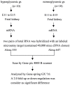
Figure 1. Schematic diagram of gene screening involved in hyperglycemia induce fetal nephropathy.
We isolated two pairs of mRNA from E11 to E15 of fetal renal tissue either in hyperglycemic or normoglycemic state, then hybridized with 40,000 labeled cDNA microarray target. Differential expression more than twofold was defined as a positive gene (group: gr.).
Figure 2
Figure 2. Serial gene expression patterns from E11 to E15 of fetal renal tissue.
Measurement of serial GDNF, EGR-1, Ras, Raf, MEK and ERK gene expression by quantitative renal time PCR (qPCR) on cDNA from E11 to E15 of fetal renal tissue, either in hyperglycemic or normoglycemic state. Change pattern was very similar in these genes, all of them highly expressed at E12, dramatically dropping at E13, then progressively increasing from E14 to E15. (Fig. 2a. GDNF, 2b. EGR-1, 2c. Ras, 2d. Raf, 2e. MEK, 2f. ERK, and summary 2 g; n = 10).
Figure 3
Figure 3. Effects of high glucose on GDNF-dependent phosphorylation of cRaf, MEK and ERK protein expression.
HRPTE cells were treated with or without 20 mM glucose and/or GDNF (100 ng/ml) for 30 minutes, protein measured by Western blot. Phosphorylated cRaf, MEK and ERK significantly increased after co-treatment with high glucose and GDNF, the latter downregulated by pretreated with ERK inhibitor, PD98059 for 1 hour. Bands were quantified by densitometry, values normalized to that of β-actin protein in the same sample. Similar results emerged from three independent experiments. * vs lane 1 p<0.05, □ vs lane 2 p<0.05, # vs lane 4 p<0.05 (Fig 3a). EGR-1 mRNA expression was significantly increased in a time-dependent manner. Real-time quantitative polymerase chain reaction of EGR-1 mRNA expression in HRPTE cells, with time-dependent manner was noted in high glucose concentration (20 nM) with GDNF (100 ng/ml) for 12, 24 and 48 hours. Relative amount was compared with their own β-actin mRNA. Each point represents mean ± SD of three independent experiments. *p<0.05 vs control, #p<0.05 vs GDNF 12 hrs, □p<0.05 vs GDNF 24 hours, ★p<0.05 vs. high glucose 24 hrs, ⊚p<0.05 vs. GDNF 48 hrs, ☆p<0.05 vs. high glucose 48 hr.(Fig.3b). siRNA of EGR-1 downregulated high glucose and GDNF mediated cRaf/MEK/ERK phosphorylation.(Fig.3c). High glucose enhanced GDNF-induced DNA damage was determined by the comet assay. Higher concentration of glucose led to more damaged cells being stained (Fig.3d)
Figure 4
Figure 4. DM newborn (Day 1) mouse kidney showed nephrogenesis period between 3rd and 4th stages, revealing less demarcated cortical labyrinth and medullary rays, as well as decreased fetal glomeruli and convoluted tubules in deeper cortical and juxtamedullary regions with scattered ‘-shaped’bodies, residual advancing ampullae of ureteric buds, diminished branching of developing convolution (100×) ( Fig.4a ).
Normal newborn (Day 1) mouse kidney showed nephrogenesis period near 4th (last) stage, plus clearly discernible cortical labyrinth and medullary rays with fetal glomeruli and well-developed tubules in the deeper cortical and juxtamedullary regions, developing glomeruli (100×)(Fig.4b). TUNEL assay revealed apoptosis cells increasing in metanephric mesenchyme (dark brown dots) and collapsed nephron region in newborn kidneys of the hyperglycemic group (200×) (Fig.4c). With maternal diabetes circumstances, large amounts of nephrons collapsed (HE stain, Fig. 4d 200× arrow, Fig. 4e 600× arrow). Day 1 hyperglycemic mice showed detachment between ureteric branch and metanephros (blue arrow), as well as collapse of nephrons (yellow arrow) (Figs.4d–4e).Serial changes of GDNF, EGR-1 and ERK-2 expression on branching morphogenesis: Upper parts were fetal kidney tissue from hyperglycemic versus lower parts from normoglycemic mothers. At E12, week expression of EGR-1, GDNF and ERK-2 in intermediate mesoderm of fetal kidney tissue from hyperglycemic mothers (Figs.3f, 3h and 3j). Ureteric buds unable to invade metanephric mesenchyme correlated with reduced immunoreactivity in the tubular cell basement membrane of fetal kidney tissue from hyperglycemic mother on E15 (Figs. 3g, 3i and 3k, 600×; similar results noted in six independent experiments) (Figs.4f–k).

References

    1. Eriksson UJ (1995) The pathogenesis of congenital malformations in diabetic pregnancy. Diabetes Metab Rev 11: 63–82. - PubMed
    1. Suhoene L, Hiilesmaa V, Termo K (2000) Glycaemic control during early pregnancy and fetal malformations in women with type I diabetes mellitus. Diabetologia 43: 79–82. - PubMed
    1. Mills Jl (1982) Malformations in infants of diabetic mothers. Teratology 25: 385–94. - PMC - PubMed
    1. Kitzmiller JL, Gavin LA, Gin Gd, Jovanovic-Peterson L, Main EK, et al. (1991) Preconception care of diabetes: Glycemic control prevents congenital anomalies. JAMA 265: 731–736. - PubMed
    1. Lynch Sa, Wright C (1997) Sirenomelia, limb reduction defects, cardiovascular malformation, renal agenesis in an infant born to a diabetic mother. Clin Dysmorphol 6: 75–80. - PubMed

Publication types

MeSH terms

Substances