. 2013;8(2):e57828.
doi: 10.1371/journal.pone.0057828. Epub 2013 Feb 28.

Treatment with 670 nm light up regulates cytochrome C oxidase expression and reduces inflammation in an age-related macular degeneration model

Affiliations

Treatment with 670 nm light up regulates cytochrome C oxidase expression and reduces inflammation in an age-related macular degeneration model

Rana Begum et al. PLoS One. 2013.

Abstract

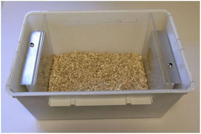
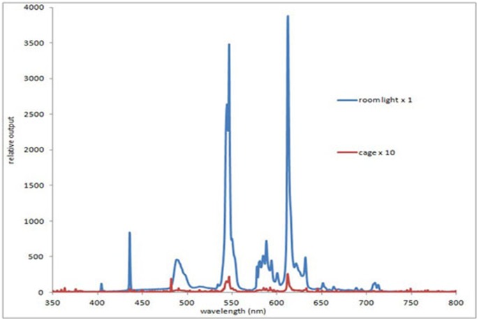
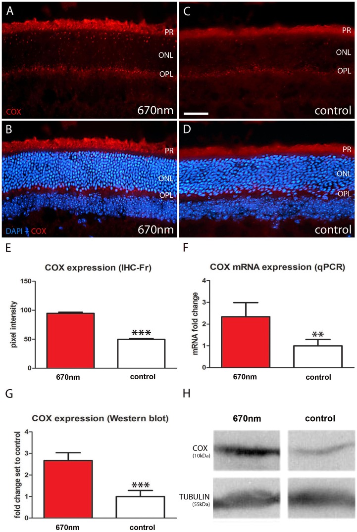
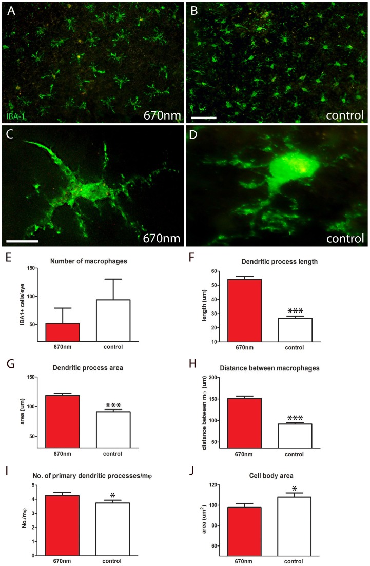
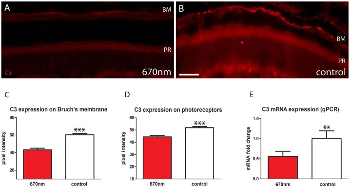
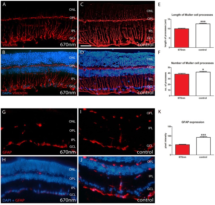
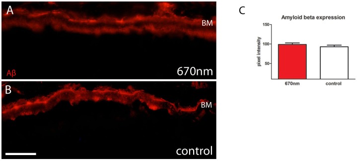
Inflammation is an umbrella feature of ageing. It is present in the aged retina and many retinal diseases including age-related macular degeneration (AMD). In ageing and in AMD mitochondrial function declines. In normal ageing this can be manipulated by brief exposure to 670 nm light on the retina, which increases mitochondrial membrane potential and reduces inflammation. Here we ask if 670 nm exposure has the same ability in an aged mouse model of AMD, the complement factor H knockout (CFH(-/-)) where inflammation is a key feature. Further, we ask whether this occurs when 670 nm is delivered briefly in environmental lighting rather than directly focussed on the retina. Mice were exposed to 670 nm for 6 minutes twice a day for 14 days in the form of supplemented environmental light. Exposed animals had significant increase in cytochrome c oxidase (COX), which is a mitochondrial enzyme regulating oxidative phosphorylation.There was a significant reduction in complement component C3, an inflammatory marker in the outer retina. Vimetin and glial fibrillary acidic protein (GFAP) expression, which reflect retinal stress in Muller glia, were also significantly down regulated. There were also significant changes in outer retinal macrophage morphology. However, amyloid beta (Aβ) load, which also increases with age in the outer retina and is pro-inflammatory, did not change. Hence, 670 nm is effective in reducing inflammation probably via COX activation in mice with a genotype similar to that in 50% of AMD patients even when brief exposures are delivered via environmental lighting. Further, inflammation can be reduced independent of Aβ. The efficacy revealed here supports current early stage clinical trials of 670 nm in AMD patients.


Conflict of interest statement

Competing Interests: The authors have declared that no competing interests exist.

Figures

Figure 1
Figure 1. Cage for 670 nm exposure.
LED lights were placed at both ends of the cage behind perspex screens. The internal space occupied a volume of 6,422 cubic centimeters (13 cm×26 cm×19 cm). A maximum of five animals were caged together. The longest distance the animals were from the light source was approximately 13 cm and the minimum was 0.4 cm, which is the thickness of the perspex screens.
Figure 2
Figure 2. Spectral composition of room light in which mice were caged.
The spectrum was measured directly under the room light which had an opaque cover (blue line) and again in the cage (red line), which did not receive any direct room illumination. The measurements for the cage are X 10 of those for the room light. As common with room lighting, it is primarily composed of a series of discrete peaks, however none of these are close to 670 nm.
Figure 3
Figure 3. Mitochondrial cytochrome c oxidase expression is enhanced in 670 nm treated aged mice.
A–D. Retinal sections were immunostained with COX (red). This enzyme is involved in mitochondrial respiratory chain and is activated by 670 nm light. In the experimental groups COX expression is significantly up regulated (A–B) compared to controls (C–E, p = 0.0001). B, D. Showed label with DAPI as a counter stain. COX staining was largely confined to photoreceptor inner segments and outer plexiform layer. F–G. This result was confirmed with significant increases in COX expression in qPCR (p = 0.0030) and Western blots (p = 0.0004). H. Differences for tubulin between groups were non-significant. Abbreviations, cytochrome c oxidase (COX), photoreceptor (PR), outer nuclear layer (ONL) and outer plexiform layer (OPL). Scale bar A–D = 40 µm.
Figure 4
Figure 4. IBA-1 staining showed significantly different macrophage morphology between 670 nm and control groups.
A–D. RPE flat mounts labelled with IBA-1 (green) to identify macrophages. After 14 days of treatment with 670 nm light these cells had significantly altered morphology over a range of metrics. E. Number of IBA-1+ cells per eye was measured; there was a reduction in the number of macrophages following treatment, but this was not statistically significant. F,G. Dendritic process length and area were significantly increased by 670 nm light (27%, 28% respectively) following treatment (p = 0.0001 for each). H. The distance between macrophages was measured from nucleus of one cell to its nearest neighbour, which also showed a significant increase (p = 0.0001). I. Not only were the 670 nm treated cells larger they also had more primary processes (p = 0.05). J. Even though these cells had a greater dendritic field and territory they had smaller cell bodies in comparison to controls (p = 0.05). Abbreviations, retinal pigmented epithelial (RPE), ionized calcium-binding adaptor molecule 1 (IBA-1), macrophages (mφ). Scale bars A, B = 40 µm, C,D = 20 µm.
Figure 5
Figure 5. Outer retinal inflammation is significantly reduced following 670 nm treatment.
A.B. Retinal sections stained with C3 (red). This accumulates on Bruch’s membrane and outer segments. C.D Following 670 nm treatment C3 was significantly reduced on Bruch’s membrane and photoreceptor outer segments (p = 0.0001 for each). E. These data were confirmed with qPCR analysis, which showed again a statistically significant reduction in C3 expression following treatment (p = 0.0031). Abbreviations, Bruch’s membrane (BM), photoreceptor (PR), complement component (C3). Scale bars = 40 µm.
Figure 6
Figure 6. Retinal stress is significantly reduced following 670 nm treatment.
Vimentin and GFAP are Muller cell markers, which are up regulated in ageing and when the retina is damaged or stressed. A–D. 670 nm treated groups showed significantly reduced vimentin labelling both in terms of length of Muller cells processes (E, p = 0.0001) and their number (F, p = 0.0329). G–J. Shows similar differences for GFAP with a statistically significant reduction following 670 nm treatment (K, p = 0.0001). In both A–D and G–J upper panels are immunostaining alone (red) while in lower panels DAPI is shown as a counter stain (blue). Abbreviations, outer nuclear layer (ONL), outer plexiform layer (OPL), inner plexiform layer (IPL), ganglion cell layer (GCL) and glial fibrillary acidic protein (GFAP). Scale bar A–D = 40 µm.
Figure 7
Figure 7. Amyloid beta expression is unaffected by 670 nm treatment.
A.B. Aβ (red) expression was measured at the Bruch’s membrane/RPE interface and this was unchanged between experimental and control groups as shown in C. Abbreviation, amyloid beta (Aβ), Bruch’s membrane (BM), retinal pigmented epithelium (RPE). Scale bar A–B = 40 µm.

References

    1. Alonso-Fernández P, De la Fuente M (2011) Role of the immune system in aging and longevity. Curr Aging Sci 4: 78–100. - PubMed
    1. Hoh Kam J, Lenassi E, Jeffery G (2010) Viewing ageing eyes: diverse sites of amyloid Beta accumulation in the ageing mouse retina and the up-regulation of macrophages. PLoS One 5: e13127. - PMC - PubMed
    1. Anderson DH, Radeke MJ, Gallo NB, Chapin EA, Johnson PT, et al. (2010) The pivotal role of the complement system in aging and age-related macular degeneration: hypothesis re-visited. Prog Retin Eye Res 29: 95–112. - PMC - PubMed
    1. Graymore CN, Kissun RD (1969) Use of phenazine methosulphate (PMS) in the histochemical localization of lactic acid dehydrogenase (LDH) in the retina. Exp Eye Res 8: 375–8. - PubMed
    1. Barot M, Gokulgandhi MR, Mitra AK (2011) Mitochondrial dysfunction in retinal diseases. Curr Eye Res 36: 1069–77. - PMC - PubMed

Publication types

MeSH terms

Substances